
Image
|
Figure Caption
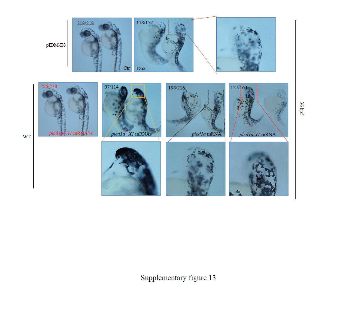
Fig. S13
Characterization of line pIDM-E8. Pictures of WT and mutant embryos with differing treatments at 36 hpf as indicated. For mRNA injection, WT embryos were injected with plcd1a mRNA, or plcd1aX1 mRNA, or plcd1a together with plcd1aX1 mRNAs (plcd1a+X1 mRNAs), or both of the mutant mRNAs carrying an early stop codon (plcd1a+X1 mRNAMs) at the one cell stage. Enlarged pictures to show delayed eye’s development. Numbers: embryos showing the displayed phenotype versus total embryos examined are provided in the corresponding pictures.
Figure Data
Acknowledgments
This image is the copyrighted work of the attributed author or publisher, and
ZFIN has permission only to display this image to its users.
Additional permissions should be obtained from the applicable author or publisher of the image.
Full text @ Sci. Rep.