
|
Fig. S6
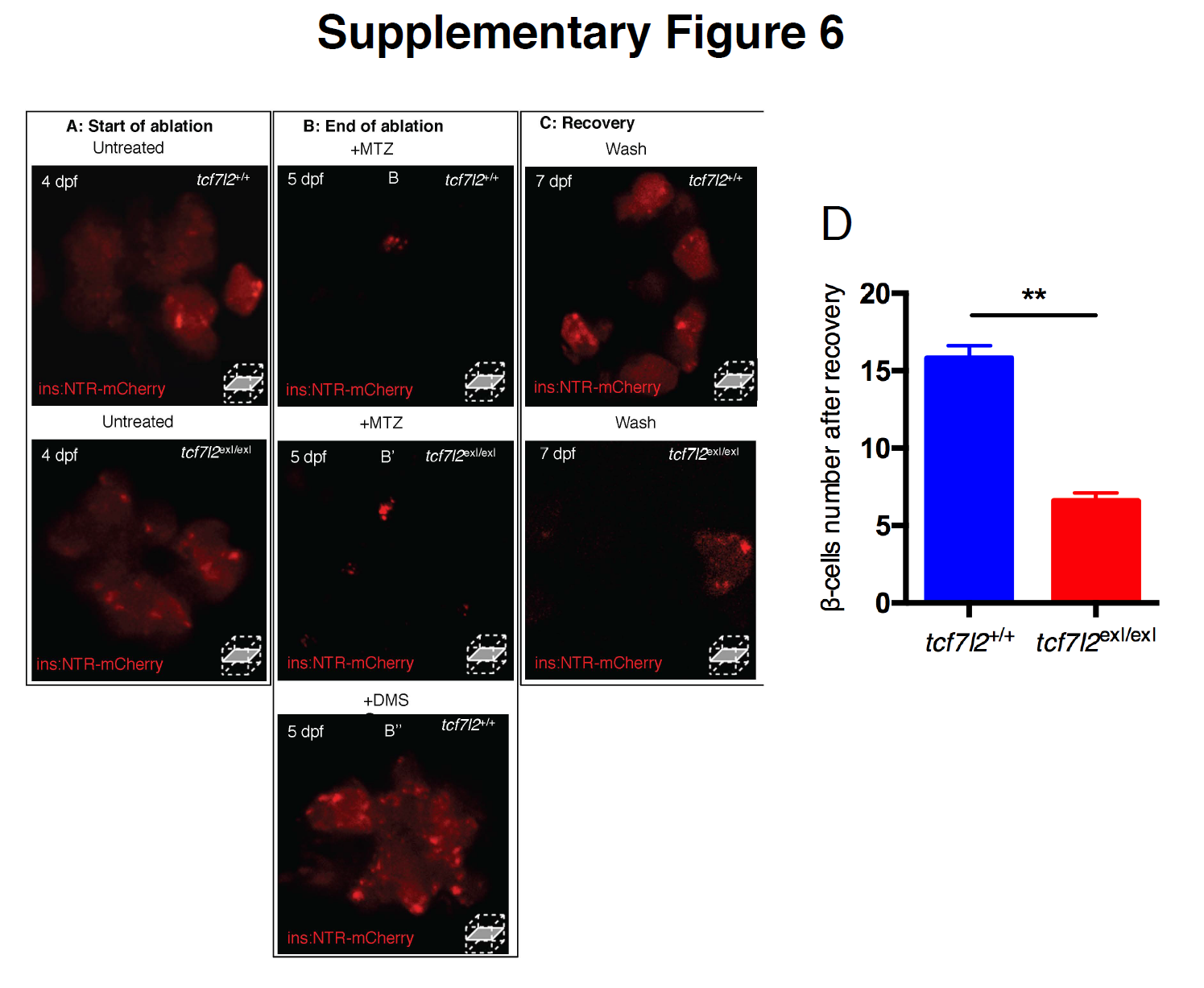
Impaired recovery of pancreatic β cells in tcf7l2exI/exI mutants. Confocal microscopy was used to monitor the progression of ablation in tcf7l2exI/exI and wt in Tg(ins:NTR-mCherry) larvae throughout their treatment with DMSO or Mtz. Control and mutant larvae at 4 dpf before treatment (A), at 5 dpf, after treatment for 24 h (B) with 7 mM Mtz (B,B’) or DMSO (B’’), and at 7 dpf, after 48 h recovery (C). Loss of mCherry in treated individuals indicates that β cells have been successfully ablated; (C) fluorescence levels indicate cell recovery in the wt but not in the tcf7l2exI/exI mutant. (D) Quantification of the number of β cells after recovery of tcf7l2exI/exI and control siblings. Data were obtained from six individuals per genotype. All reference to phenotypes was confirmed by genotyping. Values represent the mean ± SEM. Asterisk above column indicate statistical differences among groups ** p<0.01.