
|
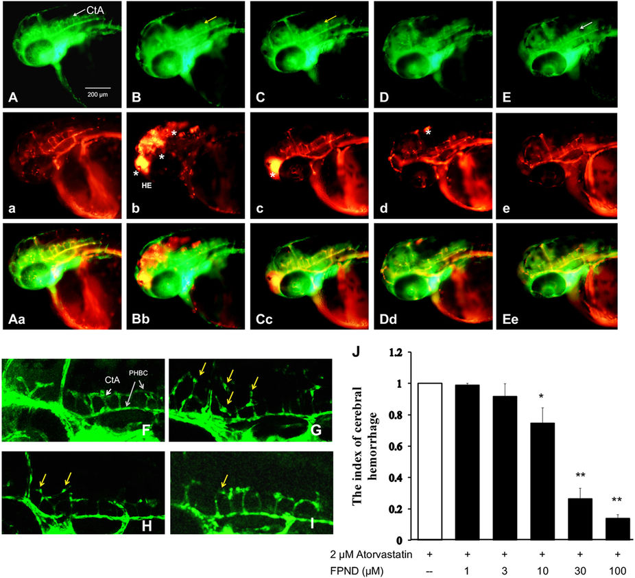
Fig. 3
The preventive effect of FPND against atorvastatin-induced cerebral hemorrhage in developing zebrafish. The 22 dpf embryos were pretreated with either 0.1% DMSO (A, B, F and G), 10 (C and H), 30 (D and I) or 100 μM (E) FPND for 3 h and replaced with 0.1% DMSO (A and F) or 2 μM atorvastatin (C–E and G–I) for 24 h. (A and F) The embryos treated with 0.2% DMSO (solvent) served as the normal control group. At 48 hpf, a lateral view of the hindbrain of the wild-type embryo shows CtA (white arrows) draining into the PHBC. Homozygous double transgenic zebrafish Tg (fli1a: EGFP) y1 and Tg (gata1: dsRed) sd2; the green fluorescence is Tg (fli1a: EGFP) y1 (a–e), the red fluorescence is Tg (gata1: dsRed) sd2 (A–E), and the third column is the overlapping photo of the first two columns (Aa, Bb, Cc, Dd and Ee). The asterisks indicate erythrocyte accumulation in the cerebral hemorrhage region of the zebrafish head. The yellow arrows indicate the morphologically abnormal blood vessels. White scale bar=200 μm. (J) The representative index of hemorrhage indicates that FPND could prevent atorvastatin-induced cerebral hemorrhage in zebrafish in a dose-dependent manner. Data presented in the bar graphs are the mean±S.D. of three independent experiments. *P<0.05 and **P<0.01 (versus control group) were considered significantly different.