
Fig. S3
- ID
- ZDB-IMAGE-180124-15
- Publication
- Hultin et al., 2017 - AmotL2 integrates polarity and junctional cues to modulate cell shape
- All Figures
- Figures for Hultin et al., 2017
|
Fig. S3
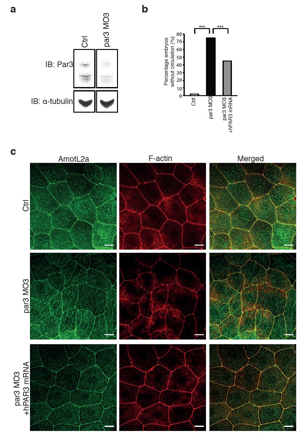
The same phenotypes of the zebrafish vasculature and skin are observed using two different par3 morpholinos. (a) Western blot showing the knock-down efficiency using par3 MO3. α-tubulin was used to control for equal loading. (b) Quantification of the circulation defect in the par3 MO3 embryos. The phenotype could be partially rescued by co-injecting the morpholino with a human PAR3 mRNA. N(ctrl)= 100 embryos, N(par3 MO)= 104 embryos, N(par3 MO+ hPAR3 mRNA)= 124 embryos. *** P≤0.001. (c) Immunofluorescence images showing AmotL2a and F-actin staining of the epidermis of ctrl (top panel), par3 MO3 (mid panel) and par3 MO3 + hPAR3 mRNA (lower panel) injected zebrafish embryos. The structure of the actin cytoskeleton is disrupted in the par3 MO3 injected embryos, correlating with and altered cell shape. Both the cell morphology and the actin filaments could be restored co-injecting the morpholino with a human PAR3 mRNA.