
|
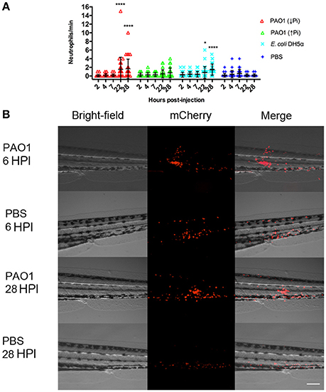
Fig. 3
Inflammation mediated by neutrophils in injected zebrafish. Tg(BACmpo:mCherry) larvae were injected at 72 HPF with 2,000–6,000 CFU of P. aeruginosa PAO1 or E. coli DH5α into the caudal artery. Sterile PBS was injected as control. (A) Larvae were injected with P. aeruginosa PAO1 grown in PGS (↓Pi) medium (red), P. aeruginosa PAO1 grown in PGS (↑Pi) medium (green), E coli DH5α (light blue) or sterile PBS medium (blue). Neutrophils that passed through the site of injection by the caudal artery in 1 min at 2, 4, 7, 22, and 28 HPI were counted. Each symbol represents a different zebrafish larva. Statistical differences with the control were determined. *P ≤0.05. ****P ≤ 0.0001. (B) Tg(BACmpo:mCherry) larvae injected at 72 hpf with P. aeruginosa grown in PGS (↓Pi) medium or sterile PBS medium were imaged at 6 and 28 HPI at the site of injection. Scale: 100 μm.