
|
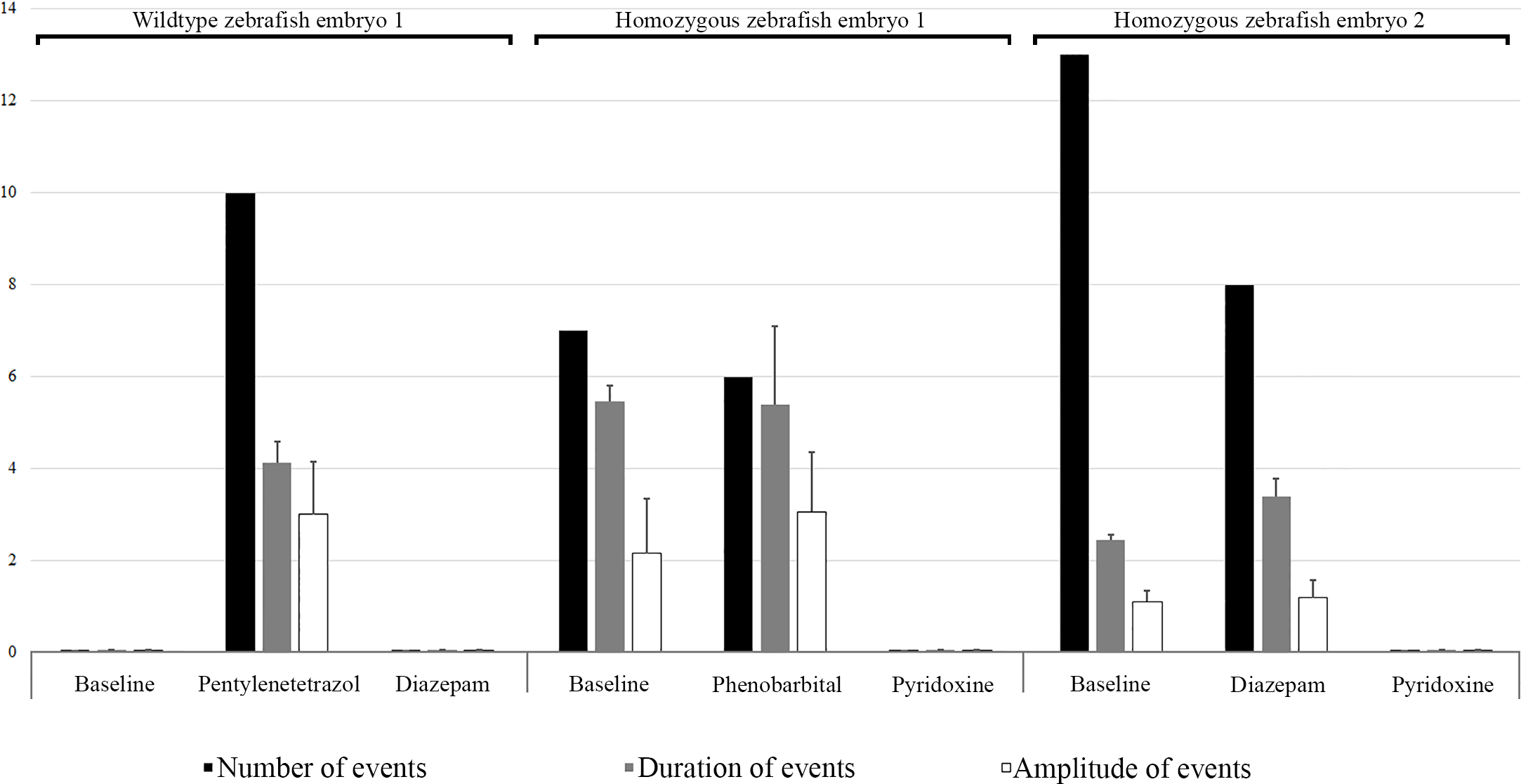
Fig. 5 EEG results of two knock-out homozygous aldh7a1 embryos and wild type embryo at 9 dpf. The results represented as number of events (per 300 seconds), duration of each event (seconds) and amplitude (mV) of each event as baseline and on treatment. Each event was considered as a single spike discharge of the EEG recording. Right panel reports events in a knock-out homozygous aldh7a1 embryo, their treatment with 100 μM diazepam and followed by 25 mM pyridoxine treatment. The middle panel shows events in another knock-out homozygous aldh7a1 embryo, their treatment with 100 μM diazepam and followed by 25 mM pyridoxine treatment. The left panel reports events in a wild type on 15 mM pentylenetetrazole to produce spikes and treat them with 100 μM diazepam. Error bars of are standard error of the mean (SEM).