
|
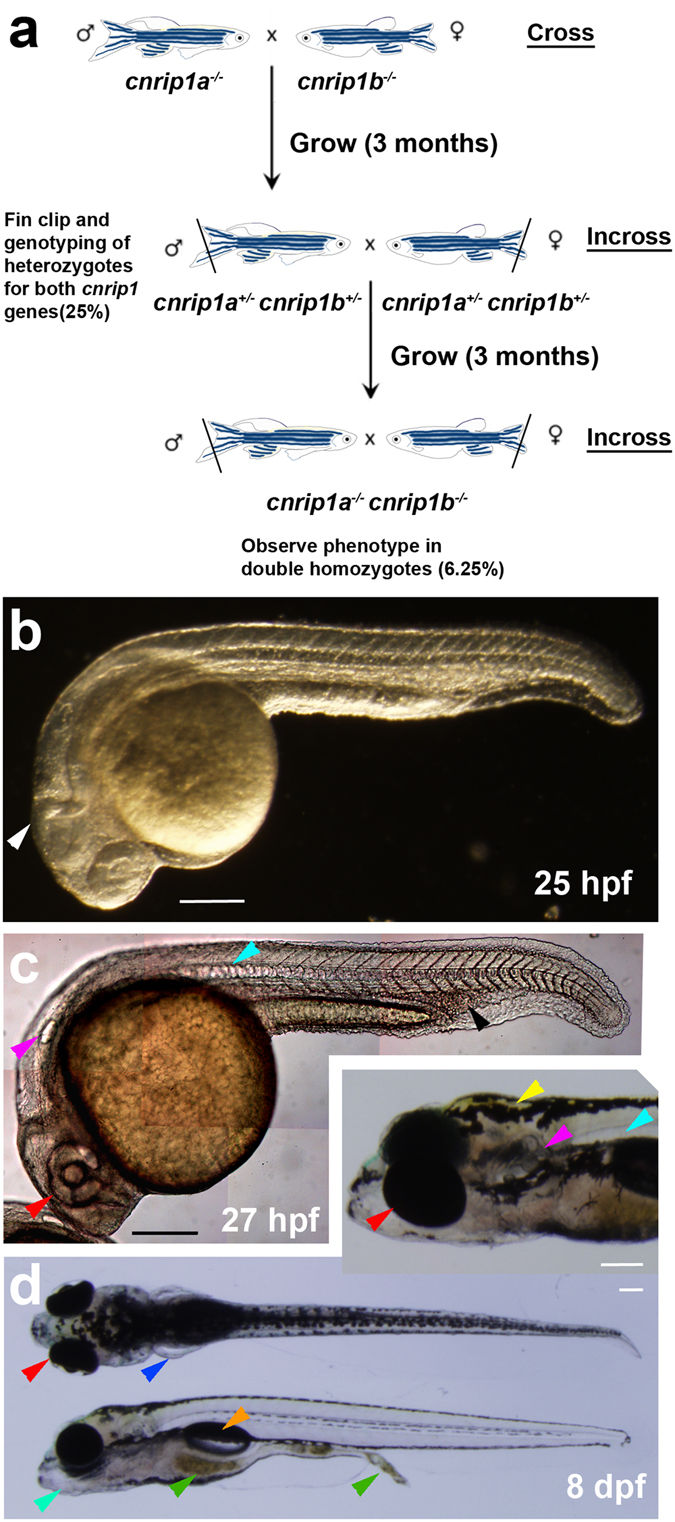
Fig. 4
Maternal zygotic double cnrip1a kg98 ;cnrip1b kg101 mutant fish develop normally. (a) Schematic of crosses to generate a cnrip1 double mutant. (b) Darkfield image of 1 dpf double mutant indicating normal complex CNS folds (white arrowhead). (c) Differential interference contrast image of 1 dpf double mutant with arrowheads indicating normal eye (red), ear (purple), notochord (cyan) and haematopoetic tissue (black). (d) Dorsal, lateral and oblique (inset) views of 8 dpf larvae with arrowheads indicating pectoral fins (blue), jaw (turquoise), food traversing gut (green), swim bladder (orange) and xanthophores (yellow). Quantitative evidence of reproducibility is given in Table S1. All fish shown with anterior to left and dorsal up. Bars = 200 μm.