
|
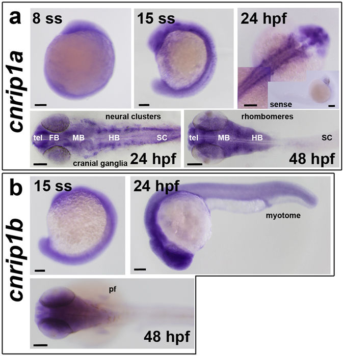
Fig. 2
Accumulation of mRNAs from cnrip1a and cnrip1b genes. Whole mount in situ mRNA hybridisation of embryos at the indicated stages for antisense probes to cnrip1a (a) and cnrip1b (b). Lateral views (first two images in each panel) are anterior to top dorsal to left, except 24 hpf in which anterior is to left and dorsal to top. Dorsal views (remaining images) are anterior to left flatmounts (a) or wholemount (b), except panel a top right, which is a wholemount montage with anterior to top right. Sense control in inset in panel a top right is anterior to right dorsal to bottom. Quantitative evidence of reproducibility is given in Table S1. tel telencephalon, FB forebrain, MB midbrain, HB hindbrain, SC spinal cord, pf pectoral fin. Bars = 100 µm.