
|
Fig. 2
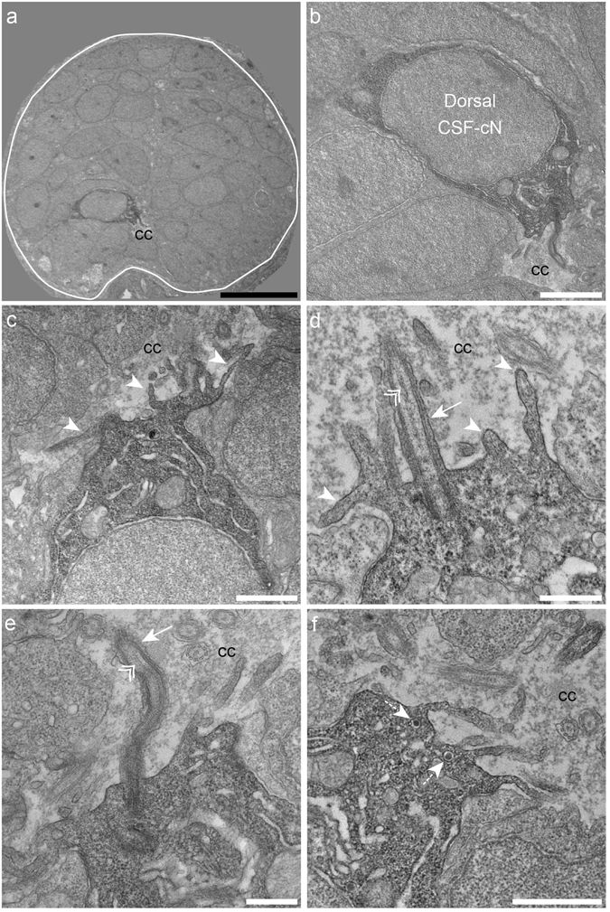
Dorsal spinal CSF-cNs also exhibit ultrastructural properties of sensory neurons. (a) Transverse section of the spinal cord showing restricted deposition of DAB in a dorsal CSF-cN. (b) Overall view of a DAB+ dorsal CSF-cN contacting the central canal (cc). (c,d) Dorsal CSF-cNs bear at the apical pole multiple microvilli (arrowheads). (d,e) In the apical pole is located a cilium (arrow) with two central microtubule singlets along the axoneme (double arrowhead), reminiscent of a motile cilium. (f) Dorsal CSF-cN also exhibit LGV distributed in the cytoplasm (dotted arrows). Scale bar = 10 μm (a), 2 μm (b), 1 µm (c,f) and 500 nm (d,e).