
|
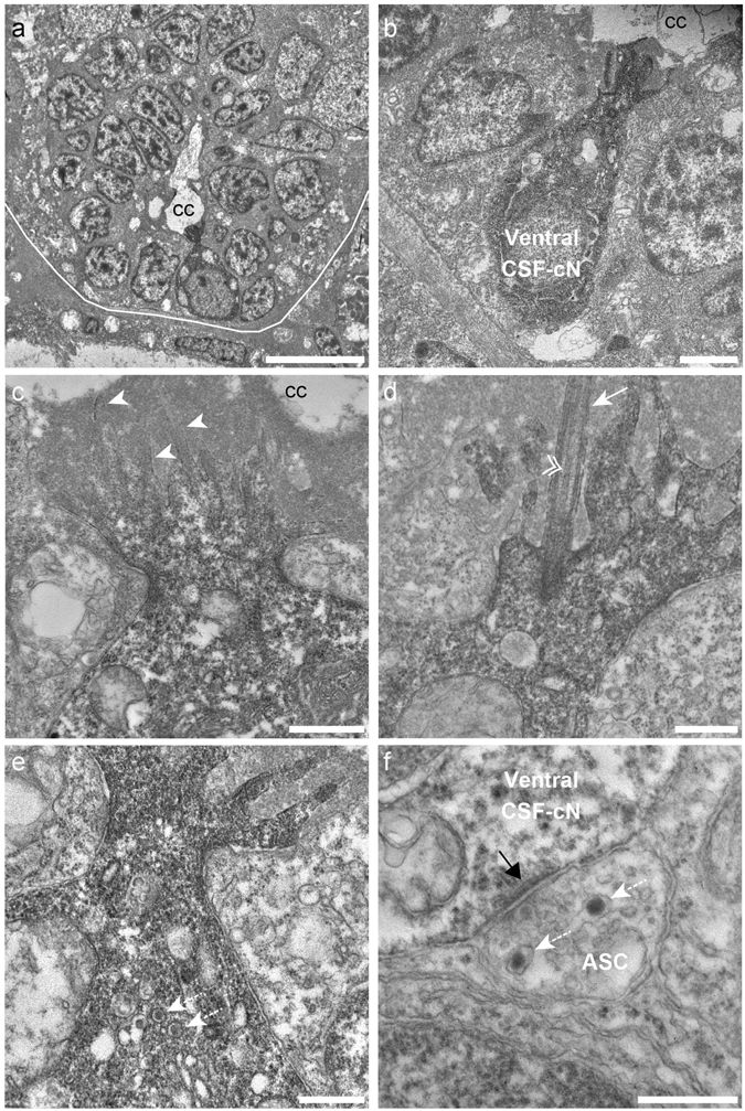
Fig. 1
Ventral CSF-cNs exhibit an apical extension composed of microvilli and a kinocilium in the spinal cord. (a) Transverse section of the spinal cord showing restricted deposition of DAB in a ventral CSF-cN. (b) Overall view of a DAB+ ventral CSF-cN contacting the central canal (cc) and surrounded by ependymal cells. (c) Ventral CSF-cNs project at the apical pole an extension toward the central canal bearing several microvilli (arrowheads). (d) Within this extension is located a cilium (arrows) with two central microtubule along the axoneme (double arrowhead), suggesting a motile cilium. Large granular vesicles (LGV) are observed in the cytoplasm (e, dotted arrows) and axo-somatic synaptic contacts in the basal pole (f, ASC). Note the symmetry of the synaptic contact (black arrow) is reminiscent of an inhibitory synapse. Note the presence of LGV in the axon (f, dotted arrows). Scale bar = 10 μm (a), 2 µm (b), 1 μm (c), 500 nm (d,e) and 400 nm (f).