
|
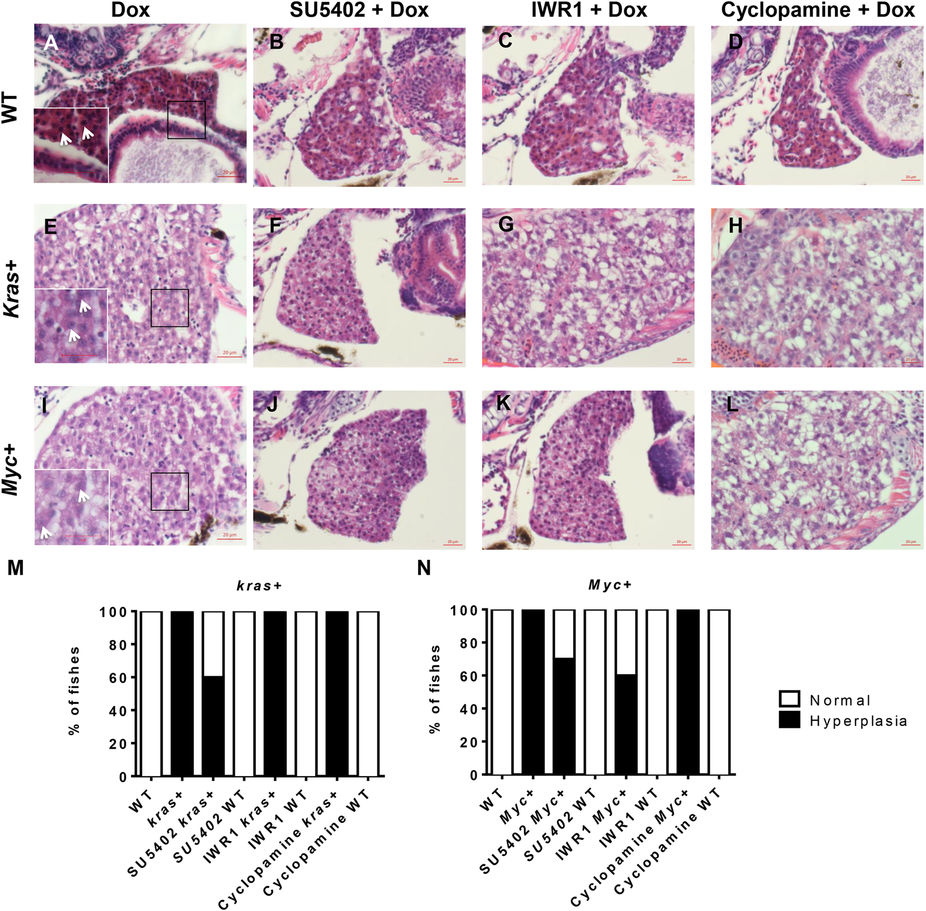
Fig. 6
Histological examination of krasV12- and Myc-induced carcinogenesis.
7 dpf WT, kras+ and Myc+ larvae were treated with 10 μM SU5402, 10 μM IWR1 or 10 μM cyclopamine in the presence of 10 μg/ml Dox, and subjected histological analysis. (A–D) Representative liver images of 7 dpf WT larvae. Inset in (A) is a magnified area in the box with arrows pointing nucleoli. (E–H) Representative liver images of 7 dpf kras+ larvae. Inset in (E) is a magnified area in the box with arrows pointing to nucleoli of condensed nuclei. (I–L) Representative liver images of 7 dpf Myc+ liver larvae. Inset in (I) is a magnified area in the box with arrows pointing to nucleoli of condensed nuclei. (M) Quantification of liver histology observed for kras+ larvae. (N) Quantification of liver histology observed for Myc+ larvae. N = 10 from each group; scale bar = 20 μm.