
|
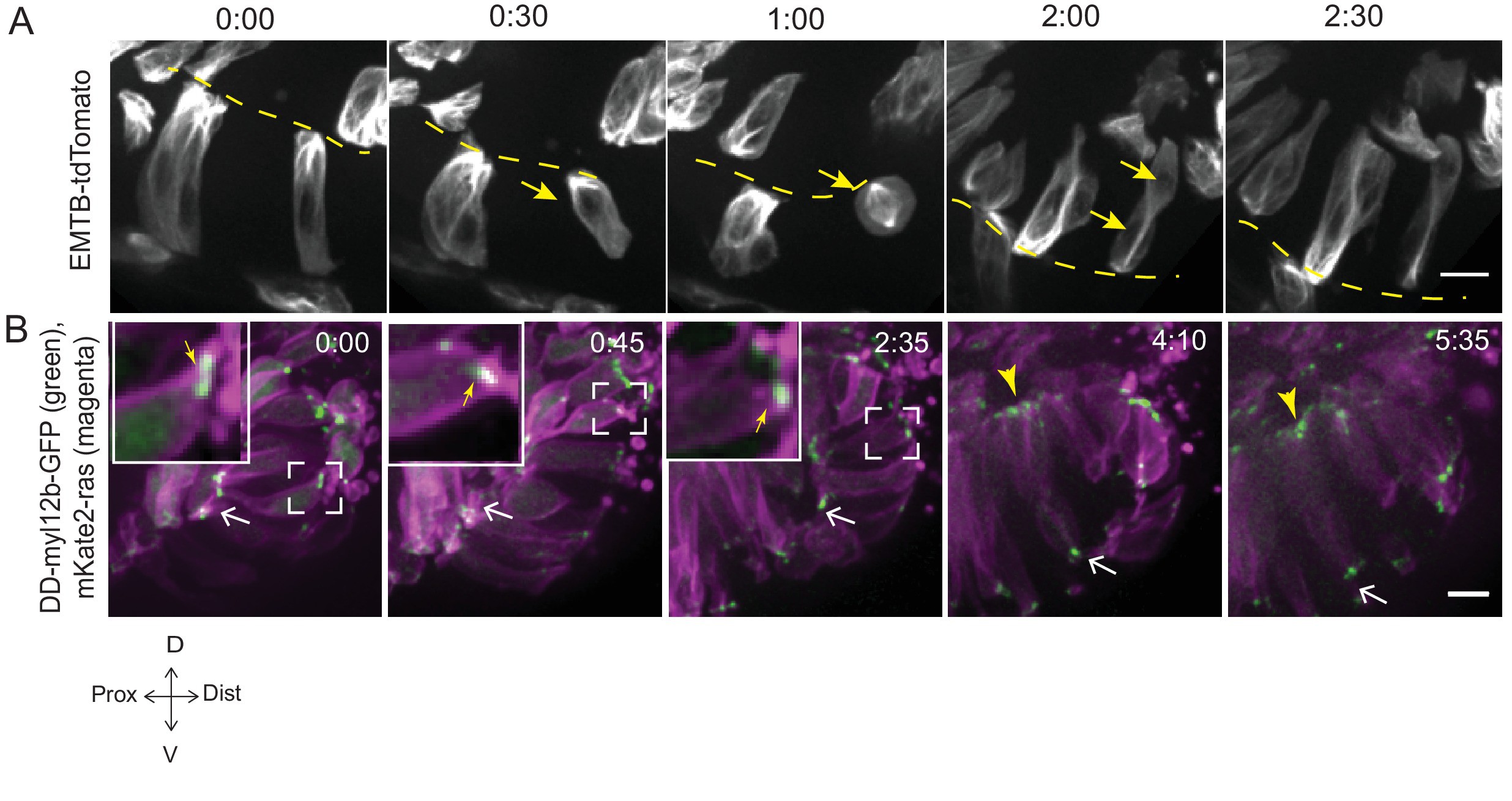
Fig. 3 S1
Microtubule and myosin dynamics during rim involution.
(A) Time-lapse imaging of rim zone with mosaic expression of EMTB-tdTomato.Arrows mark the cell dividing during rim involution. Dashed line marks the apical side. Imaging started at 17–18 ss. N = 4. (B) Time-lapse imaging of rim zone with mosaic expression of DD-myl12b-GFP and mKate2-ras. Inlays show zoomed marked area. Yellow arrows indicate basal myosin punctae in the rim cells. White arrows mark adherens junctions. Arrowheads mark basally enriched stable myosin pool in the RNE. Frames from Video 9. Imaging started at 15–16 ss. N = 5. Scale bars = 10 µm, Time h:min.