
Image
|
Figure Caption
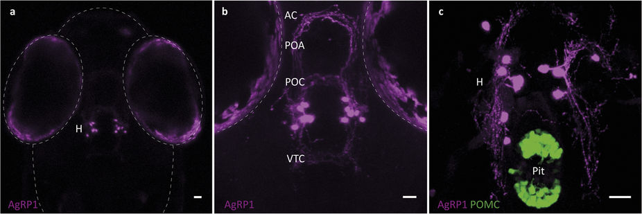
Fig. 4
Anatomical organization of AgRP1 neurons.
(a) Dorsal view of a 5-dpf agrp1:mCherry larva, showing AgRP1 somata localized at the ventral periventricular hypothalamus (H). (b) Higher magnification of the hypothalamus reveals AgRP1 projections towards the rostral, intermediate and dorsal hypothalamus, the preoptic area (POA), the anterior commissure (AC), the post-optic commissure (POC) and the ventral tegmental commissure (VTC). (c) Ventral view of a 8-dpf agrp1:mCherry; Tg(pomc:EGFP)zf44 larva shows that the AgRP1 neurons (magenta) do not project to the pituitary (Pit, green). Scale bar, 25 μm.
Figure Data
Acknowledgments
This image is the copyrighted work of the attributed author or publisher, and
ZFIN has permission only to display this image to its users.
Additional permissions should be obtained from the applicable author or publisher of the image.
Full text @ Sci. Rep.