
|
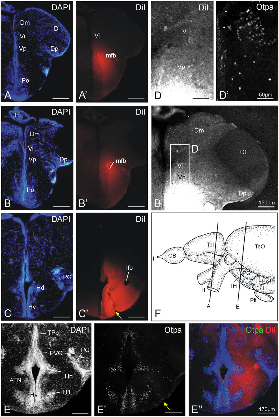
Fig. 4
Neuronal connections after a unilateral DiI injection into the tuberal hypothalamus in adult zebrafish shown at three levels from anterior (A,A') to posterior (C,C'; note yellow arrow at injection site) with corresponding DAPI and fluorescent photomicrographs demonstrating tracing results. (B'') Confocal photomicrograph shows retrograde tracing result in the telencephalon at the level of the intermediate nucleus of the ventral telencephalon (Vi). Note also that Dm, but not Dl, has retrogradely labeled cells (see text). (D,D') shows confocal blow-up of B'' (D) and corresponding Otpa stain (D'). (E–E'') details another injection site (yellow arrow) which is shown for DAPI, Otpa and DiI in confocal photomicrographs. (F) shows section levels of (A) and (E). Section (B) is immediately caudal to (A), and section (C) is at the same level as (E). Abbreviations: ATN: anterior tuberal nucleus; Dm: medial zone of dorsal telencephalic area; Dl: lateral zone of dorsal telencephalic area; Dp: posterior zone of dorsal telencephalic area; OB: olfactory bulb; E: epiphysis (pineal); ENv: ventral entopeduncular nucleus; Hd: dorsal zone of periventricular hypothalamus; Hv: ventral zone of periventricular hypothalamus; lfb: lateral forebrain bundle; LH: lateral hypothalamic nucleus; LI: hypothalamic lobus inferior; lot: lateral olfactory tract; mfb: medial forebrain bundle; mot: medial olfactory tract; PG: preglomerular complex; Pit: pituitary; Po: preoptic region; PPa: anterior parvocellular preoptic nucleus; PVO: paraventricular organ; Vd: dorsal nucleus of ventral telencephalic area; SY: sulcus ypsiloniformis; TeO: optic tectum; TH: tuberal hypothalamus; TLa: torus lateralis; TPp: periventricular part of posterior tuberculum; Vi: imtermediate nucleus of ventral telencephalon; Vp: posterior nucleus of ventral telencephalic area; Vv: ventral nucleus of ventral telencephalic area. I: olfactory nerve; II: optic nerve.