
|
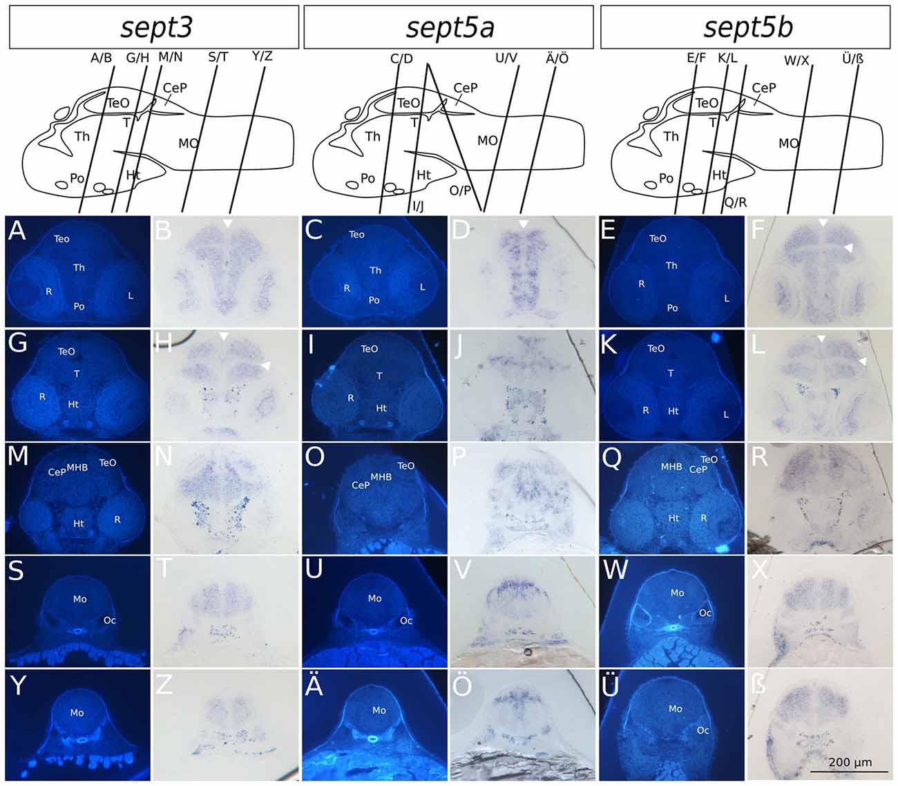
Fig. 4
Transverse sections through 2 dpf zebrafish larvae stained with ISH probes against sept3, sept5a and sept5b. 2 dpf zebrafish larvae were stained for sept3 (A,B,G,H,M,N,S,T,Y,Z), sept5a (C,D,I,J,O,P,U,V,Ä,Ö) and sept5b (E,F,K,L,Q,R,W,X,Ü,ß), embedded in epon, and cut in 8 μm thick section. The schemes (modified from Mueller and Wullimann, 2015) are showing the sagittal views of a 2 dpf zebrafish brain with indicated section planes. For better orientation the ISH sections were counterstained with Hoechst (A,G,M,S,Y,C,I,O,U,Ä,E,K,Q,W,Ü). Septin expression is indicated by the blue color (B,H,N,T,Z,D,J,P,V,Ö,F,L,R,X,ß). The retina (R), tectum opticum (TeO), tegmentum (T), preoptic region (Po), thalamus (Th), hypothalamus (Ht), medulla oblongata (Mo), cerebella plate (CeP), MHB and otic capsule (Oc), are marked. The ventricles are indicated with white arrowheads. Schemes for better orientation are modified from Mueller and Wullimann (2015) 2 dpf schemes.