
|
Fig. 3
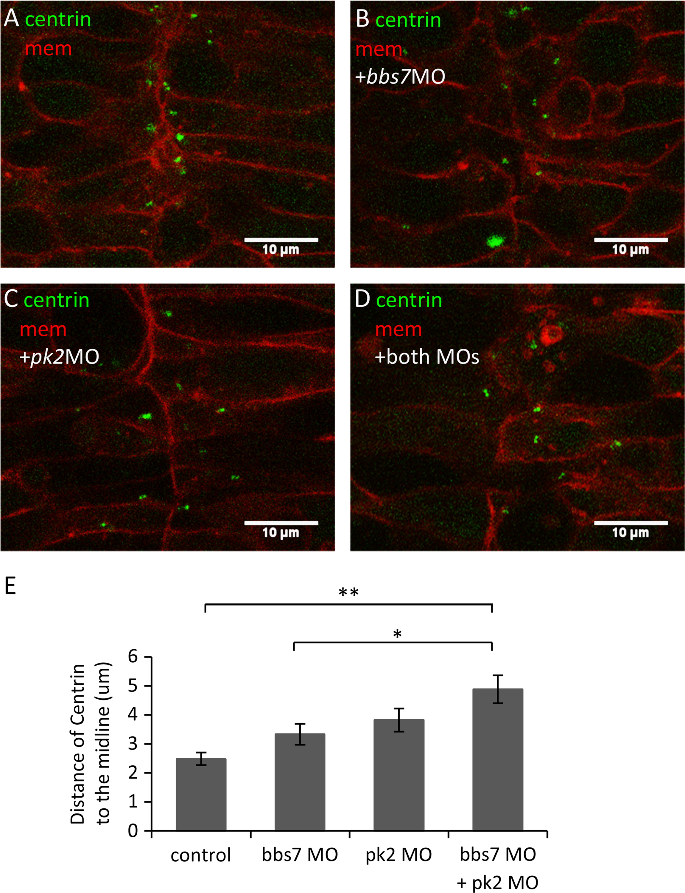
pk2 and bbs7 Double knockdown shows defects in centriole placement in the neural tube. (A)–(D) Centrin localization in the developing neural tube in pk2 and bbs7 single or double knockdown embryos. Green, Centrin-GFP; Red, mcherry-CAAX. In the control (A), centrin-GFP are localized close to the midline. In the single and double knockdown embryos ((B)–(D)) some cells have lateral placement of Centrin. Overall, the cell shape does not change. Scale bars are 10 μm in each image. (E) Quantification of distances of Centrin to the midline. While bbs7 and pk2 knockdown each leads to slight increase in the distance of Centrin to the midline, the double knockdown displays a significant increase in the distance. For control, n=119; bbs7 MO, n=33; pk2 MO, n=30; bbs7 MO+pk2 MO, n=45. Error bars are presented as standard error. **p<0.01; *p<0.05; ANOVA with Tukey. (For interpretation of the references to color in this figure legend, the reader is referred to the web version of this article.)
Reprinted from Developmental Biology, 392(2), Mei, X., Westfall, T.A., Zhang, Q., Sheffield, V.C., Bassuk, A.G., Slusarski, D.C., Functional characterization of Prickle2 and BBS7 identify overlapping phenotypes yet distinct mechanisms, 245-55, Copyright (2014) with permission from Elsevier. Full text @ Dev. Biol.