
Fig. 1
|
Fig. 1
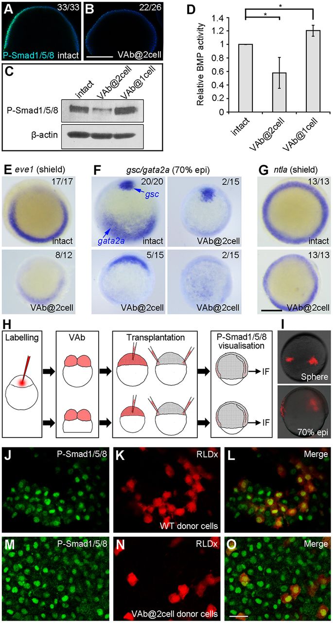
VAb@2cell inhibits BMP signalling. (A,B) Decreased P-Smad1/5/8 immunostaining (green), merged with DAPI (blue), in VAb@2cell embryos at 60% epiboly. (C,D) Analysis by western blotting of P-Smad1/5/8 levels in VAb@2cell and VAb@1cell embryos at 70% epiboly. P-Smad1/5/8 levels are normalised to β-actin, and BMP activity in intact embryos is set as 1. Data are mean±s.d. from three independent experiments. (E-G) Analysis by in situ hybridisation of indicated genes. Animal pole views with dorsal at the top. (H) Procedure of cell transplantation to visualise cell non-autonomous inhibition of BMP signalling in VAb@2cell embryos using P-Smad1/5/8 immunofluorescence (IF) staining. (I) RLDx-labelled donor cells transplanted at different locations of the margin in unlabelled intact embryos at 70% epiboly. (J-O) Similar presence of P-Smad1/5/8 immunostaining in transplanted cells derived from intact (J-L) or VAb@2cell (M-O) embryos. Scale bars: 250 µm in A,B,E-G; 20 µm in J-O.