
|
Fig. 2
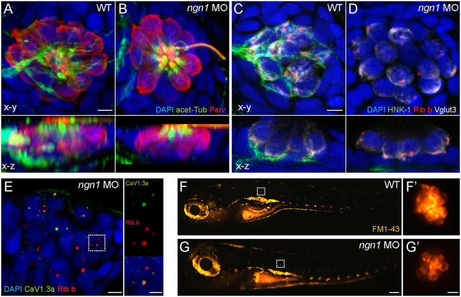
Characterization of ngn11 morphant NM hair cells.
(A,B) Representative max intensity top-down (x-y) and side-view (x-z) projections of imunolabeled Parvalbumin (red) labeling hair cells, acetylated-Tubulin (green) labeling neurons and the apical-region of hair cells, and DAPI labeling nuclei in posterior LL NM2 of WT (A) or ngn1 MO (B) 5 dpf zebrafish larvae. Note the absence of immunolabeled green neurites beneath hair cells in the ngn1 morphant NM (x-z) image. Scale bar: 3 μm (C,D) Representative immunoabeled (x-y) and (x-z) cross-sections of a WT (A) or ngn1 MO(A’) anterior NM. Both synaptic ribbons (Ribeye; red) and synaptic vesicles (Vglut3; white) localize to the basolateral end of ngn1 MO hair cells, despite the lack of afferent innervation (HNK-1; green). Scale bar: 3 μm (E) Max-intensity(x-y) projection of imunolabeled synaptic ribbons (red) and the presynaptic calcium channel CaV1.3a (green) in NM3 of a 5 dpf ngn1 morphant larva. Insets show colocalization of Ribeye and CaV1.3, which is a hallmark of hair-cell maturation. Scale bars: 3 μm, 1 μm (insets) (F,G) Representative lateral views of 5 dpf WT (F) or ngn1 morphant (G) larvae briefly exposed FM1-43. Insets show higher magnification top-down views of the NMs indicated by the white dashed squares. Labeling with FM1-43 indicates functional mechanotransduction machinery in ngn1 morphant hair cells.