
|
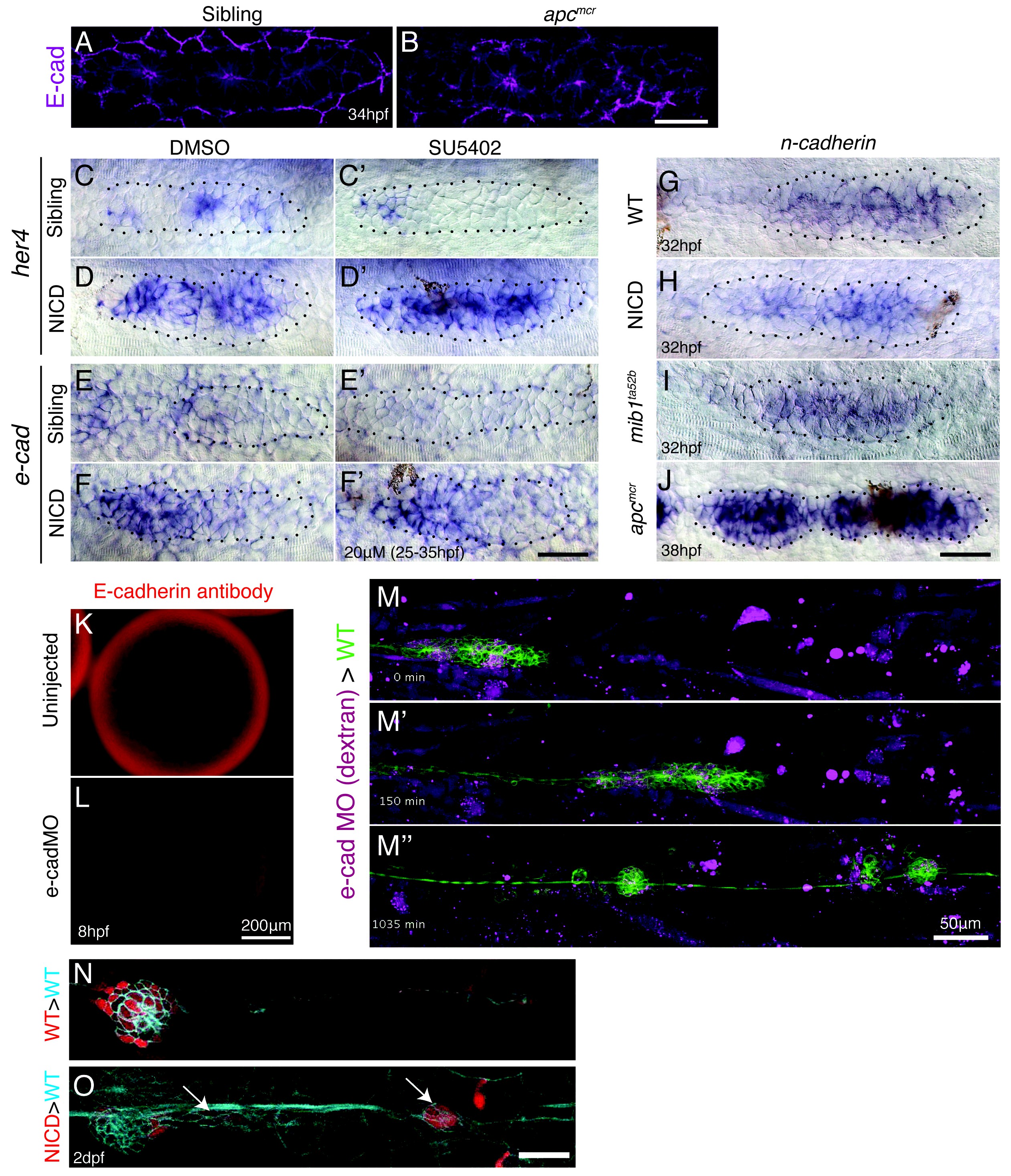
Fig. 7 S1
E-cadherin defficiency does not disrupt proneuromast formation.
(A and B) Wnt signaling upregulates E-cadherin antibody expression in the posterior lateral line primordium. Scale bar is 50 μm. (C–F') Notch regulates e-cadherin expression downstream of Fgf signaling. (C and C') The Fgfr1 inhibitor SU5402 depletes the expression of the Notch target gene her4 in the primordium. (D and D') NICD induces her4 in the absence of Fgf signaling. (E and E') Fgf is required to induce e-cadherin in the primordium. (F and F') (F) In NICD primordia, e-cadherin is upregulated, even in the absence of Fgf signaling (F'), therefore, e-cadherin is a Notch target downstream of Fgf signaling. (G–J) n-cadherin expression is unchanged in NICD and mib1ta52b primordia (I). n-cadherin is induced by Wnt signaling (J). (K–L) Injection of the translation blocking e-cadherin morpholino significantly reduces E-cadherin antibody expression in a 8 hpf embryo (L). (M–M'') Still images taken at different time points of a timelapse recording of a wildtype primordium in green that contains transplanted e-cadherin morpholino injected cells. Transplantation of e-cadherin morpholino injected cells into a wildtype host does not disrupt proneuromast formation. (O) Transplanted NICD cells form clusters (marked with the white arrows), which is not observed when wildtype cells (N) are transplanted into a wildtype host (quantification in Figure 7K). All scale bars are 25 μm, unless stated otherwise.