
|
Fig. 1 S1
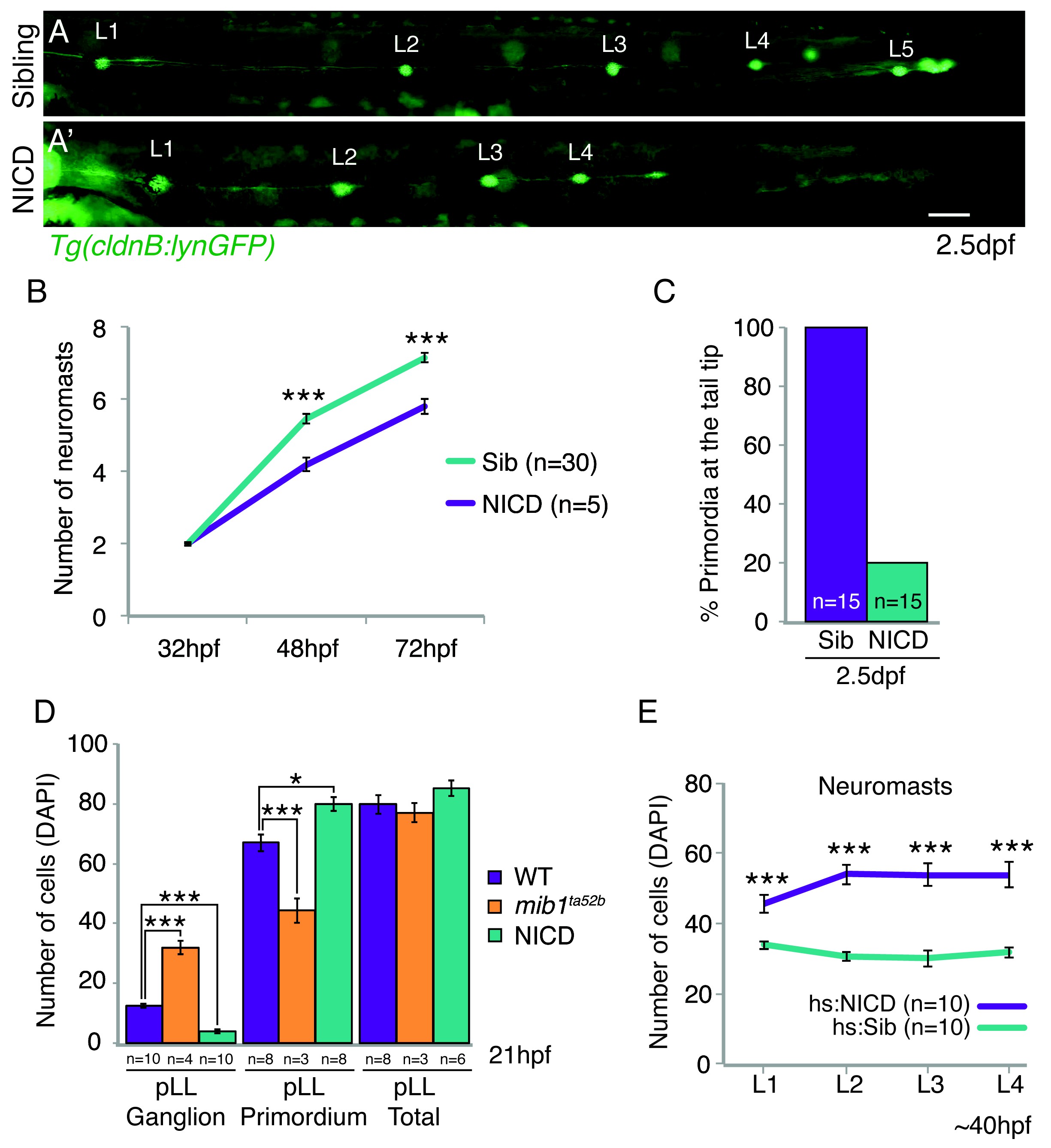
NICD primordia do not migrate to the tail tip and deposit fewer neuromasts.
(A and A') posterior lateral line in 2.5 dpf Tg(cldnB:lynGFP) larvae. Scale bar is 100 μm. (A' and B) NICD primordia deposit fewer neuromasts. Error bars represent standard error (p<0.001=*** Student's t test). (C) 80% of NICD primordia stall and fail to reach the tail tip. (D) Notch signaling regulates posterior lateral line primordium fate versus ganglion fate (also in [Mizoguchi et al., 2011]). The NICD posterior lateral line (pLL) primordium has significantly more cells compared to wildtype at the expense of ganglion cells. In contrast, in mib1ta52b mutants more cells are specified towards the ganglion fate and less cells contribute to the primordium. Error bars represent one-way ANOVA with Tukey pairwise comparison test (p<0.05=*, p<0.001=***). (E) Induction of Notch after the primordium and ganglion have separated leads to a significant increase in the number of cells in neuromasts (L1–4) compared to a heat-shocked sibling. Notch overexpression was induced by a 39°C heat-shock for 45 min starting at 25 hpf (L1 was still a part of the primordium). Embryos were fixed after L4, L5 deposition (~40 hpf). Error bars indicate standard error between the hs:sibling and hs:NICD neuromast cell counts from one independent experiment (p<0.001=*** Student's t test).