
Image
|
Figure Caption
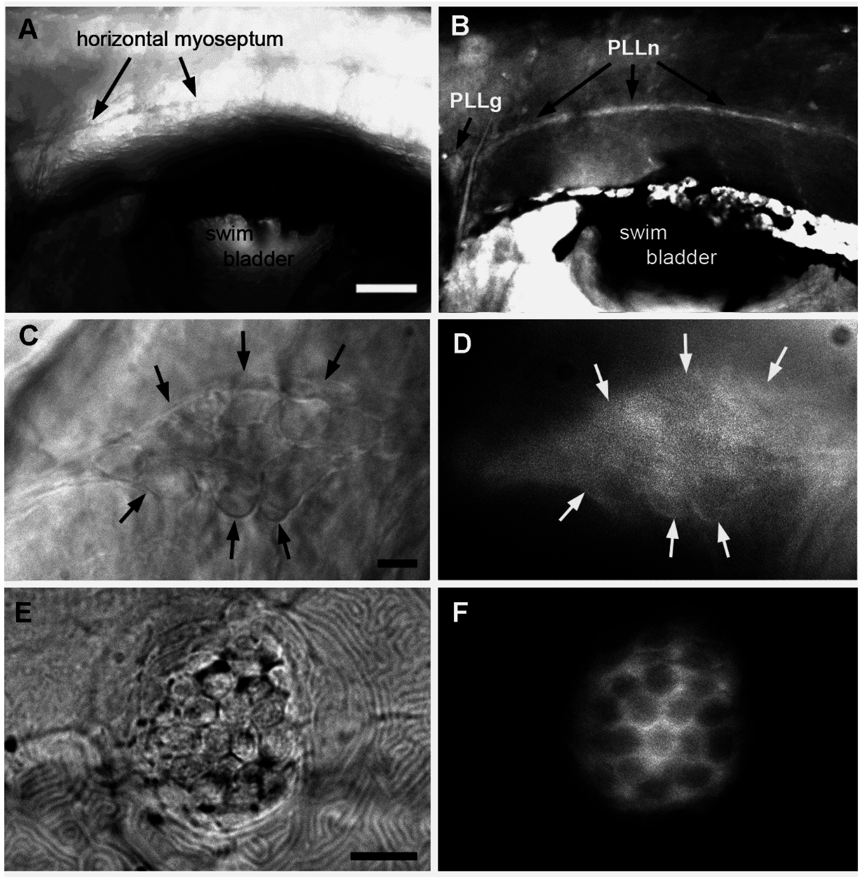
Fig. 5
Accumulation of A6 in the lateral line system of embryos that had been incubated in 50 nM A6 from 1 to 6 dpf. (A,B) Anterior trunk region; (C,D) Lateral line ganglion; (E,F) Neuromast L2; (A,C,E) Bright field; (B,D,F) Fluorescence. Arrows in (C,D) outline the PLL ganglion. Scale bars: (A) 100 μm, (C,E) 10 μm.
Acknowledgments
This image is the copyrighted work of the attributed author or publisher, and
ZFIN has permission only to display this image to its users.
Additional permissions should be obtained from the applicable author or publisher of the image.
Full text @ Int. J. Mol. Sci.