
|
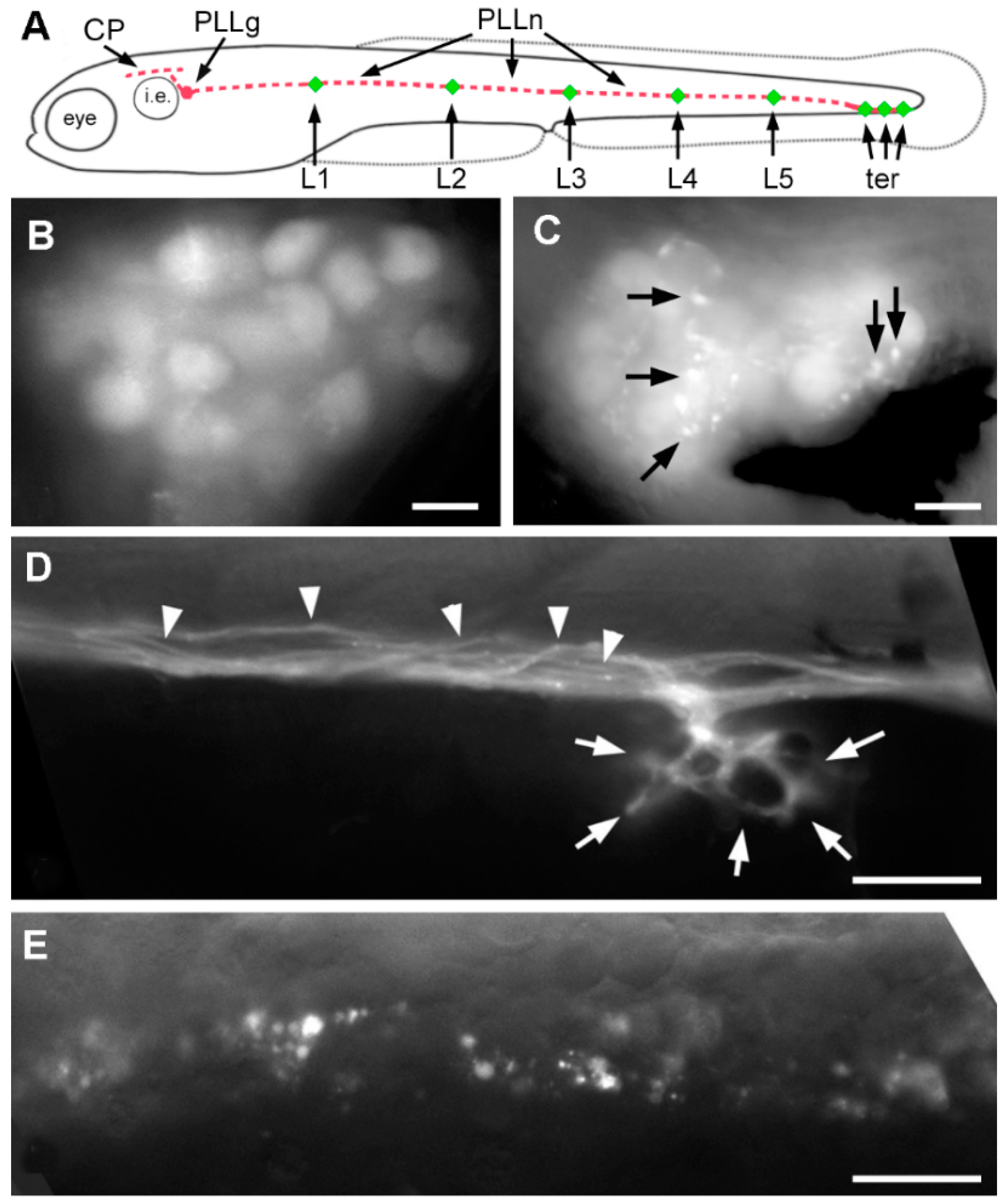
Fig. 3
Effect of A6 on the posterior lateral line (PLL) system. (A) Scheme of a 4-dpf larva showing the posterior lateral line ganglion (PLLg) just posterior to the otic vesicle (i.e., inner ear), its central projection (CP) extending along the hindbrain, and the peripheral nerve (PLLn) extending along the horizontal myoseptum towards the sensory organs (in red), neuromasts L1–L5 and terminal neuromasts, ter (in green); (B) Lateral line ganglion in the nbt-dsred line; (C) As in (B), in a larva that had been exposed to 20 µM A6 from 1 to 3 dpf; (D) Lateral line nerve and branch to a neuromast in the nbt-dsred line; (E) Lateral line nerve in a larva that had been exposed to 20 µM A6 from 1 to 3 dpf. Arrows in (C) indicate fluorescent inclusions. Arrowheads in (D) point to individual axons within the PLL nerve; arrowheads show axonal terminations surrounding the mechanosensory hair cells of a neuromast; Scale bars: 10 µm.