
Image
|
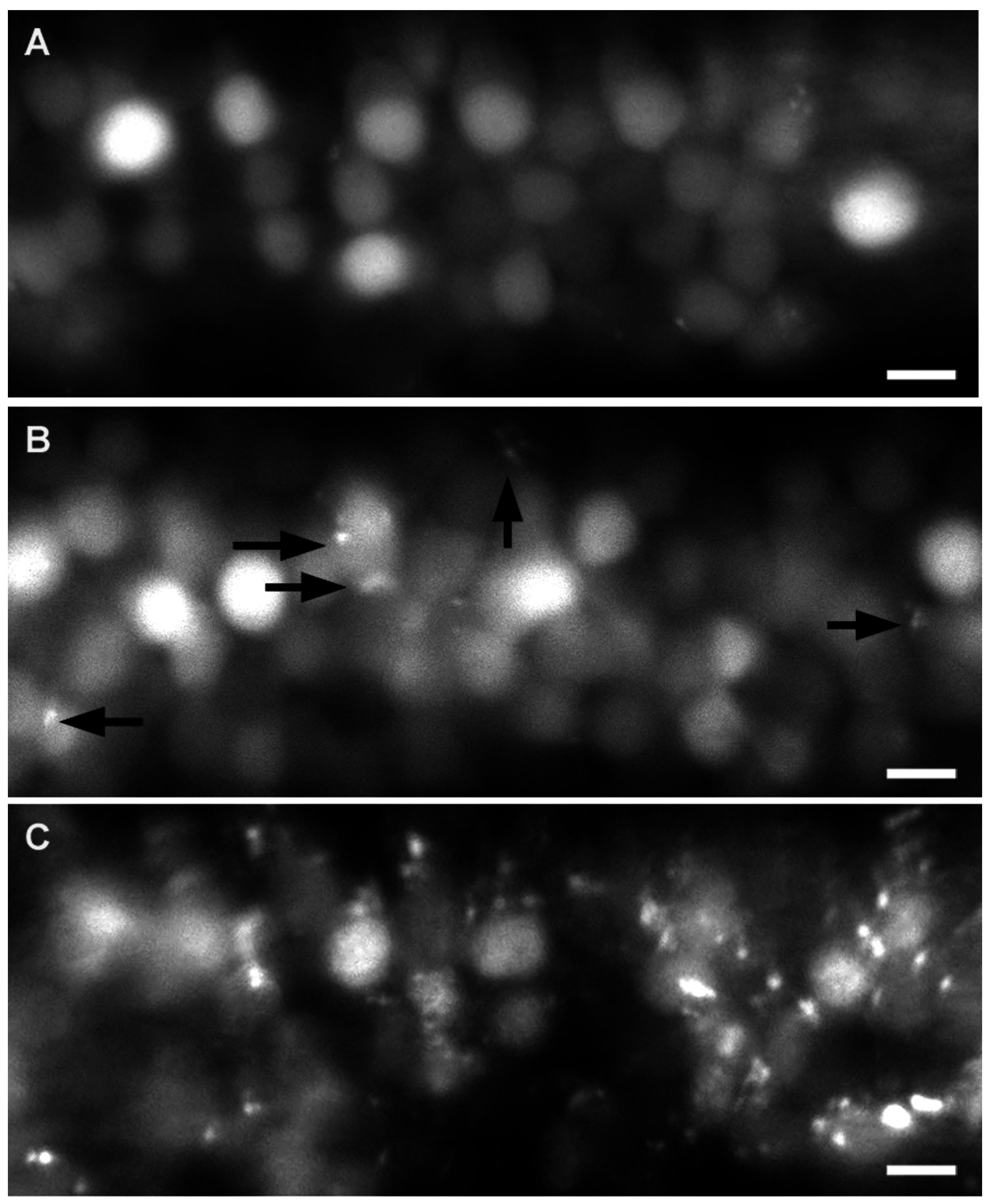
Figure Caption
Fig. 2
Effect of cyprodinyl and A6 on the central nervous system of nbt-dsred larvae. 1-dpf embryos were exposed to the solvent alone (A); to 20 µM cyprodinyl (B); or to 20 µM A6 (C), and their spinal cord neurons were visualized after two days of incubation. At least nine larvae were examined for each condition, with very similar results. Arrows indicate fluorescent inclusions. Scale bars: 10 μm.
Acknowledgments
This image is the copyrighted work of the attributed author or publisher, and
ZFIN has permission only to display this image to its users.
Additional permissions should be obtained from the applicable author or publisher of the image.
Full text @ Int. J. Mol. Sci.