
|
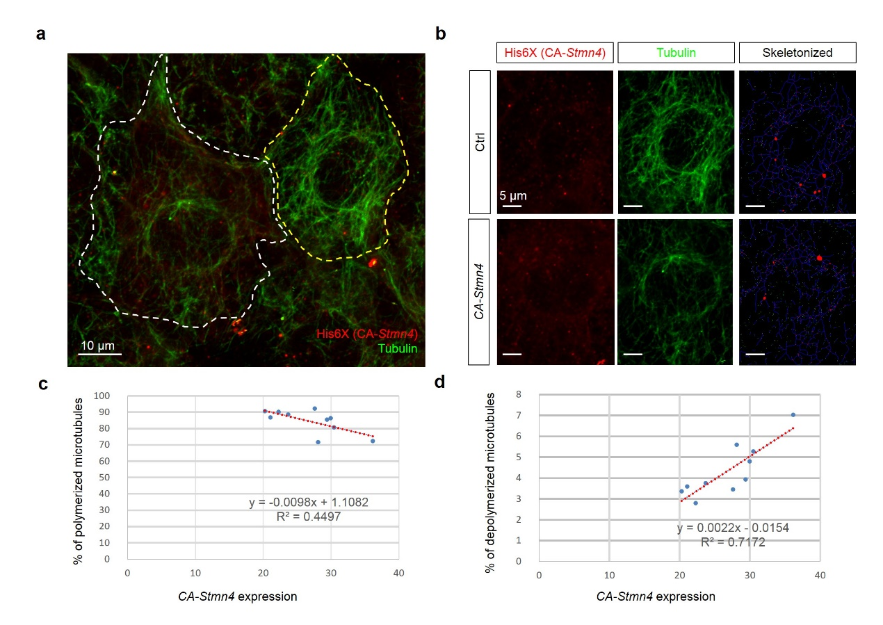
Fig. S3
Overexpression of constitutively active stmn4 induced depolymerization of microtubules in zebrafish embryos.
Zebrafish embryos were injected with plasmids containing without or with constitutively active Stmn4 (CA-Stmn4) as described in Fig. S2, cultured and fixed at the bud stage. The fixed embryos were subjected to immunohistochemistry against histidine and alpha-tubulin as described in the Materials and Methods. After staining and washing, embryos were imaged and photographed under confocal microscopy. (a) Representative image of cell overexpressed with ca-stmn4 (labeled by α-His6X, red signals) co-staining tubulin (indicated by green signals). Scale bars equal 10 μm. (b) Two separate channels one showing differential CA-Stmn4 expression level, strong (white dashed polygon, lower) and weak (yellow dashed polygon, upper), and corresponding tubulin patterns. Plugins “Skeletonized” and “AnalyzeSkeleton” of ImageJ software were utilized to transform into the right panels with labeling slabs (blue), junctions (red) and ends (green). (c,d) Percentage of polymerized or depolymerized microtubules among total tubulins were analyzed.