
|
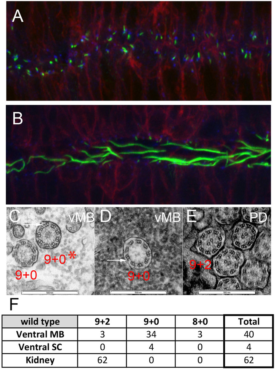
Fig. 6
Ventral midbrain and ventral spinal cord produce long cilia with 9 + 0 axonemes.
(A,B) Tg(β-actin:mGFP) embryos at 24 hpf were stained with antibodies against acetylated α-tubulin (green) and γ-tubulin (blue) to label ciliary axonemes and basal bodies, respectively. (A) Short cilia at the dorsal midbrain midline. (B) Long cilia at the ventral midbrain midline. (C–E) Wildtype embryos at 1 dpf were processed for transmission electron microscopy and analyzed in transverse sections at the levels of ventral midbrain (C,D) and pronephric duct (E). Arrows point to outer dynein arms. (F) Summary of ciliary structures in the ventral midbrain, spinal canal, and pronephric ducts of wiltype embryos.