
Image
|
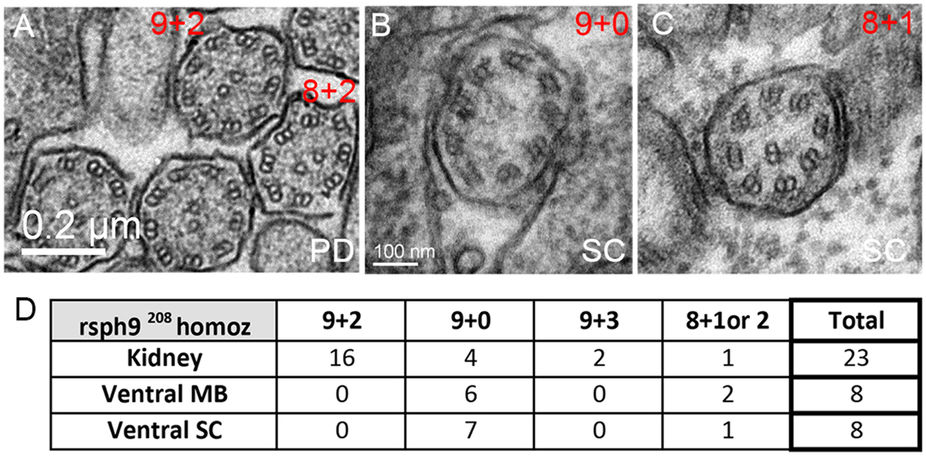
Figure Caption
Fig. 5
Ultrastructural defects in rsph9 mutant cilia.
(A–C): Representative TEM images of cilia from rsph9208/ rsph9208 embryos at 1 dpf. Both the normal 9 + 2 and aberrant 8 + 2 axonemes are present in the pronephric ducts (A). Normal 9 + 0 (B) and aberrant 8 + 1 (C) cilia are found in the ventral spinal cord. (D) Summary of ciliary structures observed by TEM in rsph9208/ rsph9208 embryos surveyed at kidney level (5 embryos, 2 experiments), midbrain level (3 embryos, 1 experiment), and spinal cord level (4 embryos, 2 experiments). PD: pronephric duct; SC: ventral spinal cord.
Figure Data
Acknowledgments
This image is the copyrighted work of the attributed author or publisher, and
ZFIN has permission only to display this image to its users.
Additional permissions should be obtained from the applicable author or publisher of the image.
Full text @ Sci. Rep.