
|
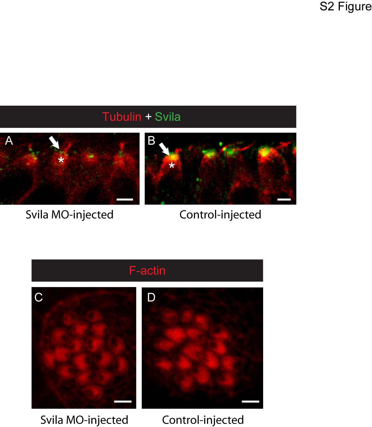
Fig. S2
Morpholino knockdown of Svila in zebrafish.
Confocal micrographs of 4-dpf zebrafish hair cells injected with a morpholino targeting Svila (A,C) or a 5-bp mismatch control morpholino (B,D). (A,B) Hair cells from the anterior macula labeled with anti-Svila (green) and anti-acetylated tubulin (red) reveal that the intensity of Svila protein at the cuticular plate (arrows) is diminished in Svila morpholino-injected fish (A) compared to fish injected with control (B), but some Svila protein is still detected (A). Fluorescence intensity of anti-Svila at the CP was compared to that associated with anti-tubulin labeling of the underlying microtubules (asterisks). Phalloidin labeling of neuromast hair cells from Svila morpholino-injected (C) and control-injected (D) fish reveals normal gross cuticular plate structure in the morphants.