
|
Fig. 3
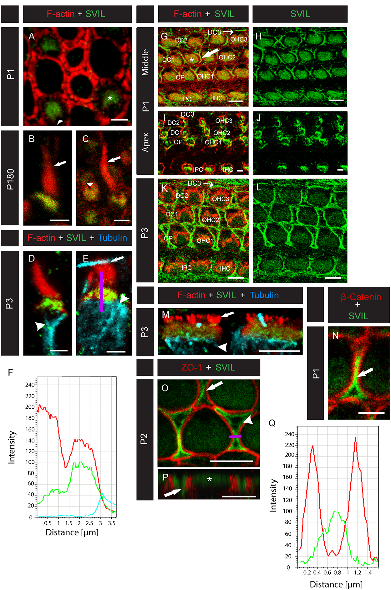
Supervillin localizes to mouse hair cell CPs and cochlear supporting cell head plates.
(A-E) Confocal micrographs of mouse vestibular hair cells labeled with anti-SVIL (green) and phalloidin (red) at different developmental stages. (A) A top-down view of several hair cells from a mouse at P1. Supervillin labels the CPs (asterisk) but not the fonticulus (arrowhead). (B,C) Views of hair cells from a 6-month-old mouse. Supervillin labels the CP but not the fonticulus (arrowhead) or stereocilia (arrows). (D,E) Type I (D) and type II (E) vestibular hair cells from a P3 mouse co-labeled with anti-tubulin (blue), which marks somatic microtubules underlying the CPs (arrowheads). Arrow in (E) indicates a kinocilium from a neighboring hair cell. A region of interest (ROI, indicated by the purple line) was selected to span the hair bundle (top portion of the line), CP (middle portion of the line), and underlying microtubules (bottom portion of the line). (F) Fluorescence intensity profile using the ROI from (E). The hair bundle (top portion of the purple ROI line) corresponds to the left region of the plot, showing robust F-actin-associated signal (red), while SVIL- (green) and tubulin-associated (blue) signals are minimal. The middle region of the plot corresponds to the CP (middle portion of the ROI line) and shows overlapping SVIL- and F-actin-associated signals; however, in the right region of the plot, only tubulin-associated signal is seen below the CP (bottom portion of the ROI line). (G-M) Confocal micrographs of mouse cochlear hair cells labeled with anti-SVIL (green) and anti-actin (red). (G-J) Hair cells at the middle (G,H) and apical (I,J) cochlear turns of a P1 mouse. At the middle turn (G,H), SVIL localizes to the CPs (asterisk) and to the region of the hair cell-supporting cell junctions (arrow). At the apical turn (I,J), SVIL co-localizes with actin near the apical surface of the developing hair cells. (K,L) In the middle turn of the P3 mouse cochlea, SVIL localizes to CPs of outer hair cells (OHCs) and inner hair cells (IHCs) and to supporting cell apicolateral margins, including those of Deiters’ cells (DC1, DC2, DC3), outer pillar cells (OP), and inner phalangeal cells (IPC). (M) Side view of two IHCs from the middle turn of a P3 mouse cochlea co-labeled with an antibody to tubulin (blue) demonstrates that SVIL localizes between the hair bundle (arrow) and somatic microtubules (arrowhead), at the region of the cuticular plate (asterisk). (N) Magnification of two OHCs and the Deiters’ cell between them (arrow) from the basal turn of a P1 mouse cochlea labeled with anti-SVIL (green) and anti-β-catenin (red). (O) Magnification of the first two rows of OHCs from the basal turn of a P2 mouse cochlea labeled with anti-SVIL (green) and anti-ZO-1 (red). SVIL strongly localizes to the apicolateral margins of the OPs (arrowhead) and the DCs (arrow). (P) Z-stacks of confocal sections were converted into a 3D model using the Leica Software. A 3D reconstruction of an OHC (asterisk) from the second row flanked by two DCs (arrow) labeled with anti-SVIL (green) and anti-ZO-1 (green) is seen. (Q) Fluorescence intensity profile of the cell in (O) using the ROI indicated by the purple line demonstrates that the signal associated with SVIL (green) is concentrated in the supporting cells, sandwiched between ZO-1-rich bands (red). In graphs in F and Q, intensity scales are linear, but the units are arbitrary. Scale bars, 2 μm.