
|
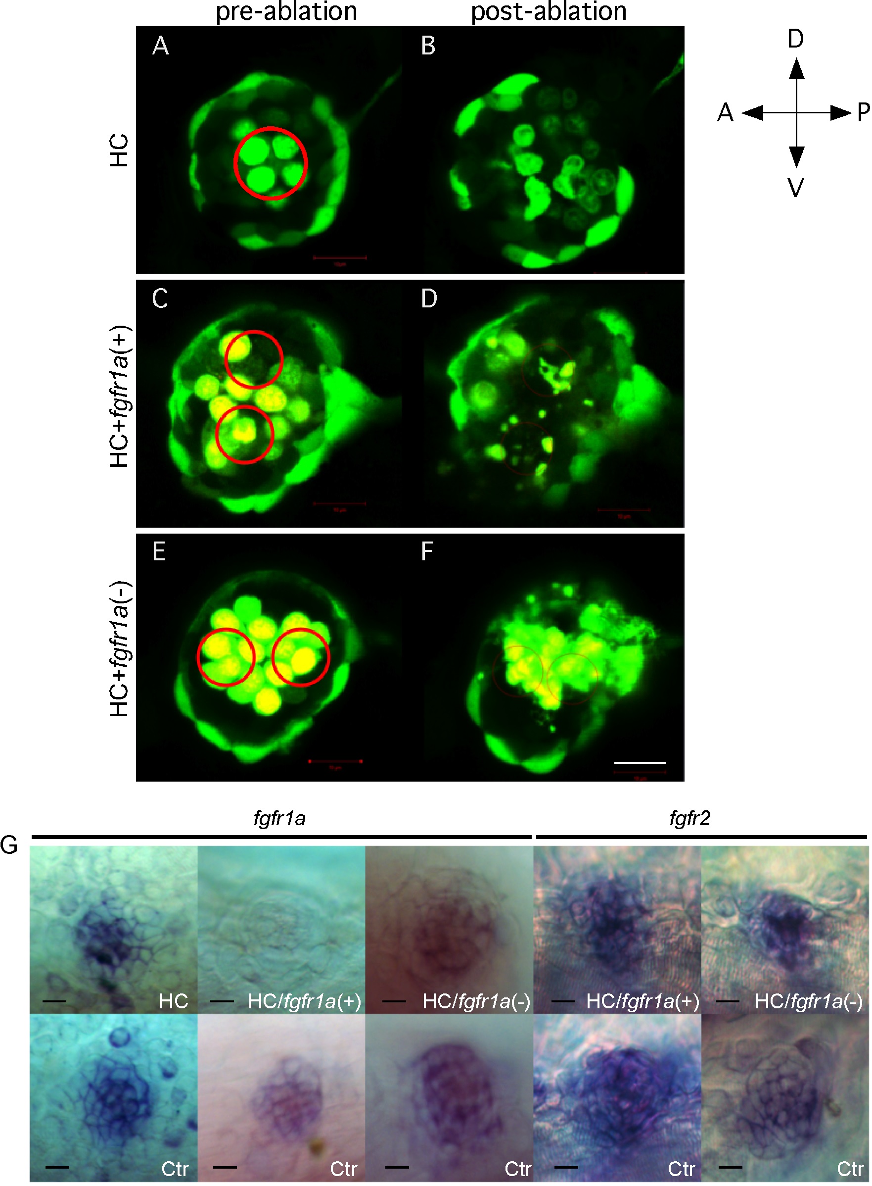
Fig. S9
Laser ablation of HCs and SCs in zebrafish neuromasts.
Hybrid larvae of pou4f3:GFP and ET20 fish were used to ablate HCs alone (A,B), and HC/fgfr1a(+) SCs (C-D), or HC/fgfr1a(-) SCs (E-F). Neuromasts before (A,C,E) and after ablation (B,D,F) were shown. The red circles are the target areas for ablation. A-P, anterior-posterior; D-V, dorsal-ventral. Scale bar, 10 μm. (G) In situ hybridization of fgfr1a confirmed the ablation of fgfr1a(+) cells in HC/fgfr1a(+) ablation group, where fgfr1a is undetectable; whereas in HCs or HC/fgfr1a(-) ablation group, fgfr1a signal is still present. Similar ablation did not change fgfr2 signal. Ctr, unablated neuromasts labeled with fgfr1a. Scale bars: 10 μm.