
|
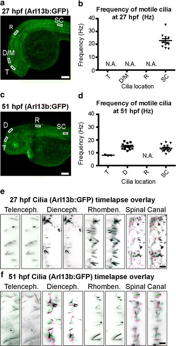
Fig. 5
Brain cilia are non-motile at early larval stage; some are motile at late larval stage. a, c Arl13b: GFP is expressed in cilia throughout the developing fish including the neuroepithelium and spinal canal. b, d Quantification of ciliary beat frequency (Hz) for motile cilia in each region at early (27 hpf) and late (51 hpf) larval stages, if any. e, f Visualization of ciliary movement for cilia in each region at 27 and 51 hpf, if any. Each of the three panels is an overlay of two sequential image acquisition frames (images were taken in the same region at distinct times at 57.22 fps, so each overlay represents a time difference of ~0.017 s). See Additional file 2: Movie 1 and Additional file 3: Movie 2 for full dataset. Colors were selected such that unmoved pixels appear black (Frame 1: green + magenta; Frame 2: red + cyan). Horizontal lines represent average vmax and error bars denote SEM. T telencephalic ventricle, D/M diencephalic/mesencephalic ventricle, R rhombencephalic ventricle, SC spinal canal ventricle analysis region, hpf hours post-fertilization, N.A. quantification of cilia frequency is not applicable because no cilia are motile. Scale bars: a, c: 100 μm; e, f: 5 μm