
|
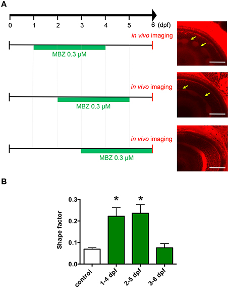
Fig. 2
Development during 2 and 3 dpf was the critical window for developmental toxicity of MBZ in the zebrafish retina. (A) Zebrafish were treated with 0.3 μM MBZ during the indicated periods. At 6 dpf, zebrafish were stained with ZMA462 and the retinas were imaged in vivo. The malformations in the IPL caused by MBZ are indicated by yellow arrows. Scale bar: 50 μm. (B) Quantitative analysis of the developmental toxicity of 0.3 μM MBZ in the zebrafish retina. The shape of the IPL in each zebrafish was quantified using the shape factor. n = 5 for control, n = 6 for 1–4 dpf, n = 4 for 2–5 dpf and 3–6 dpf, *p < 0.05 compared with control.