
|
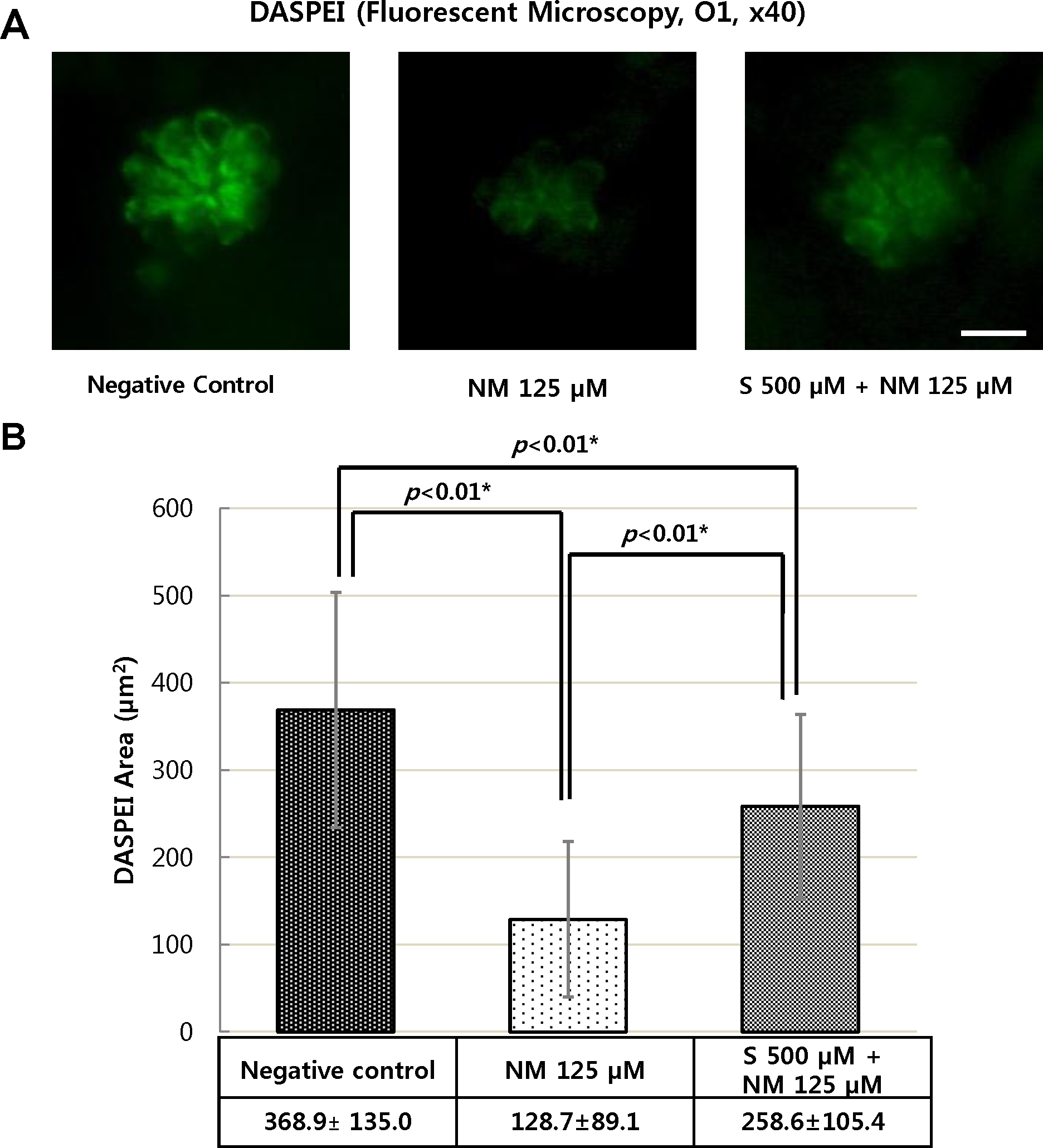
Fig. 4
Analysis of hair cell damage by DASPEI assay (×40).
The wild type zebrafish were treated with 125 μM neomycin and 500 μM sodium selenite for 1 h. Staining hair cells with DASPEI showed that treatment with 125 μM neomycin caused reduced numbers of hair cells in neuromast. However, the co-treatment with 500 μM sodium selenite protected reducing number of hair cell staining with DASPEI (A). The concentration of 500 μM of sodium selenite preserved significantly the average DASPEI area in four neuromasts (SO1, SO2, O1, and OC1) (n = 20 fish per treatment; *: statistically significant) (B). Negative control: the average DASPEI area = 368.9 ± 135.0 μm2; NM 125 μM: the average DASPEI area = 128.7 ± 89.1 μm2; S 500 μM + NM 125 μM: the average DASPEI area = 258.6 ± 105.4 μm2(B). NM, neomycin; S, sodium selenite. Scale bar = 10 μm.