
|
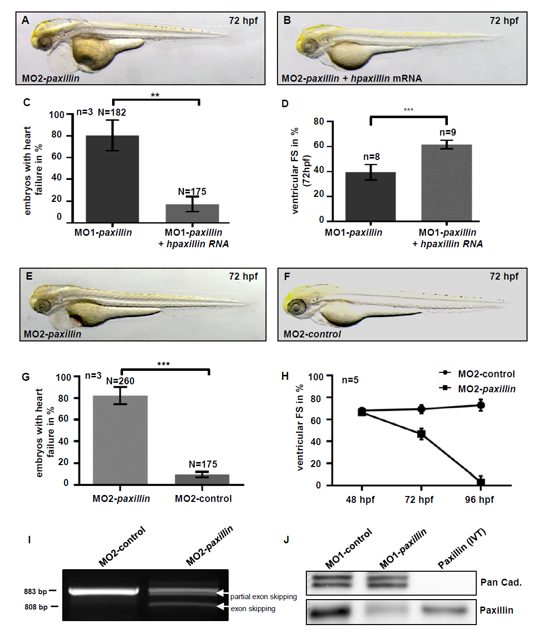
Fig. S2
Injection of human paxillin mRNA rescues the cardiac phenotype of MO1-Paxillin morphants.
(A-C) Lateral view of MO1-paxillin (A) and MO1-paxillin+human paxillin mRNA (B) injected embryos at 72 hpf. (C) Bar graphs compare average of rescued embryos after co-injection of human paxillin mRNA compared to MO1-paxillin injected embryos (n = 3; *P = 0.0073). (D) Fractional shortening (FS) measurement of rescued embryos compared to Paxillin morphant embryos at 72 hpf (n = 5). (E, F) Lateral view of a (E) 5bp-mismatch MO (MO2-control) and (F) MO2-paxillin splice MO injected embryos at 72 hpf. The heart failure phenotype of MO2-paxillin splice morphants was identical to that of embryos injected with translation blocking paxillin MO (MO1-paxillin). (G) Bar graphs compare average of affected embryos after injection of MO2-paxillin compared to 5bp-mismatch MO (MO2-control) injected embryos at 72 hpf (n = 3; *P = 0.0001). (H) FS measurements of Paxillin morphant ventricles at 48, 72 and 96 hpf (n = 5 individuals per time point). (I) RT-PCR of control- and MO2-paxillin-injected embryos. Injection of MO2-paxillin caused partial and whole skipping of exon 2 (298 bp) leading to premature termination of Paxillin protein translation. Wild-type paxillin RNA was severely reduced in Paxillin morphants (527 bp product). (J) Western blot analysis of control- (MO1-control) and MO1-paxillin-injected embryos demonstrated high efficacy of MO-mediated paxillin gene knock-down. For each sample 50 embryos were pooled and 20 μg of protein lysate were loaded per lane. In vitro translated (IVT) zebrafish Paxillin protein was used as positive control.