
|
Fig. S7
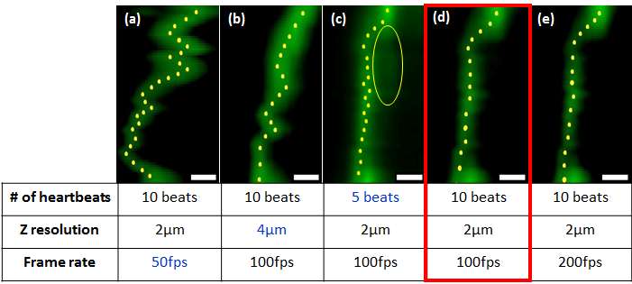
Comparison of 4-D synchronized images with a combination of 3 different parameters. (a & b) Both low frame rate and low z resolution were inadequately synchronized. Both images demonstrated a crinkled pattern in the cardiac wall. (c) Reducing the capturing number to 5 heartbeats (cardiac cycles) revealed similar synchronization with capturing 10 beats. However, negligible artifact appeared behind the wall (yellow circle). (d & e) Increasing the frame rate to 200fps revealed identical image quality to that of 100fps. Therefore, we selected (d) as the optimal combination for 4-D synchronized imaging parameters. Scale bar = 10µm.