
|
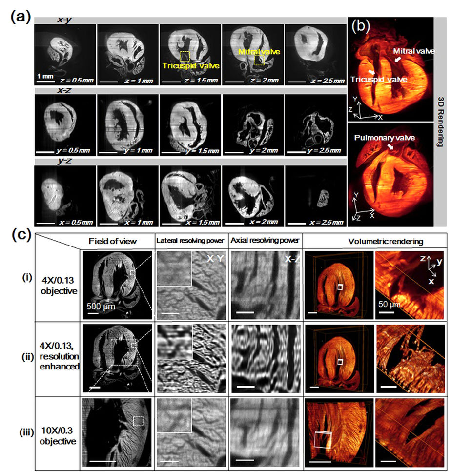
Fig. 5
Cardiac LSFM (c-LSFM) imaging of a 1-day neonate mouse heart with enhanced cellular resolution.
(a) The coronal, sagittal, and transverse planes at different depths uncover 3-D architecture. Scale bars are 1 mm in length in all of the sub-graphs. (b) The boxes were cropped from the volume rendering of the reconstructed “digital heart” to reveal the endocardial architecture. (c) The cardiac architecture is compared with the (i) 18 μm light-sheet and 4X/0.13 objective, (ii) 4X/0.13 resolution enhanced images, and (iii) 9 μm light-sheet and 10X/0.3 objective. Magnification from left to right reveals the field of view, lateral, and axial resolving power, followed by the volumetric rendering effects of 3 configurations. Myocardial orientation was resolved in detail in the resolution-enhanced c-LSFM group. All scale bars are 500 μm, except for 50 μm in the rightmost column.