
|
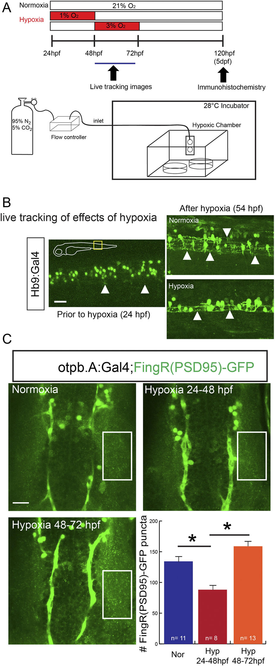
Fig. 4
FingRs reveal impairment of synapse development by developmental hypoxic injury.
(A) Schematic illustration of hypoxia exposure and imaging. (B) Demonstration of live imaging of effects of hypoxic injury on motor neuron synaptic puncta visualized by FingR(PSD)-GFP in trunk of Tg(Hb9:Gal4); Tg(FingR(PSD95)-GFP) animals. Following hypoxia there is a decrease in the number of puncta labeled by FingR(PSD95)-GFP. Confocal images, rostral to left, scale bar 10 μm. (C) Confocal images and quantification in the dopaminergic neurons in the diencephalon of Tg(otpb.A:Gal4); Tg(FingR(PSD95)-GFP) animals. Hypoxia from 24–48 hpf decreased number of puncta but later hypoxia exposure did not significantly change number; (p = 0.002; one-way ANOVA; SEM shown). Confocal z-stacks, rostral to top, scale bar 10 μm.