
|
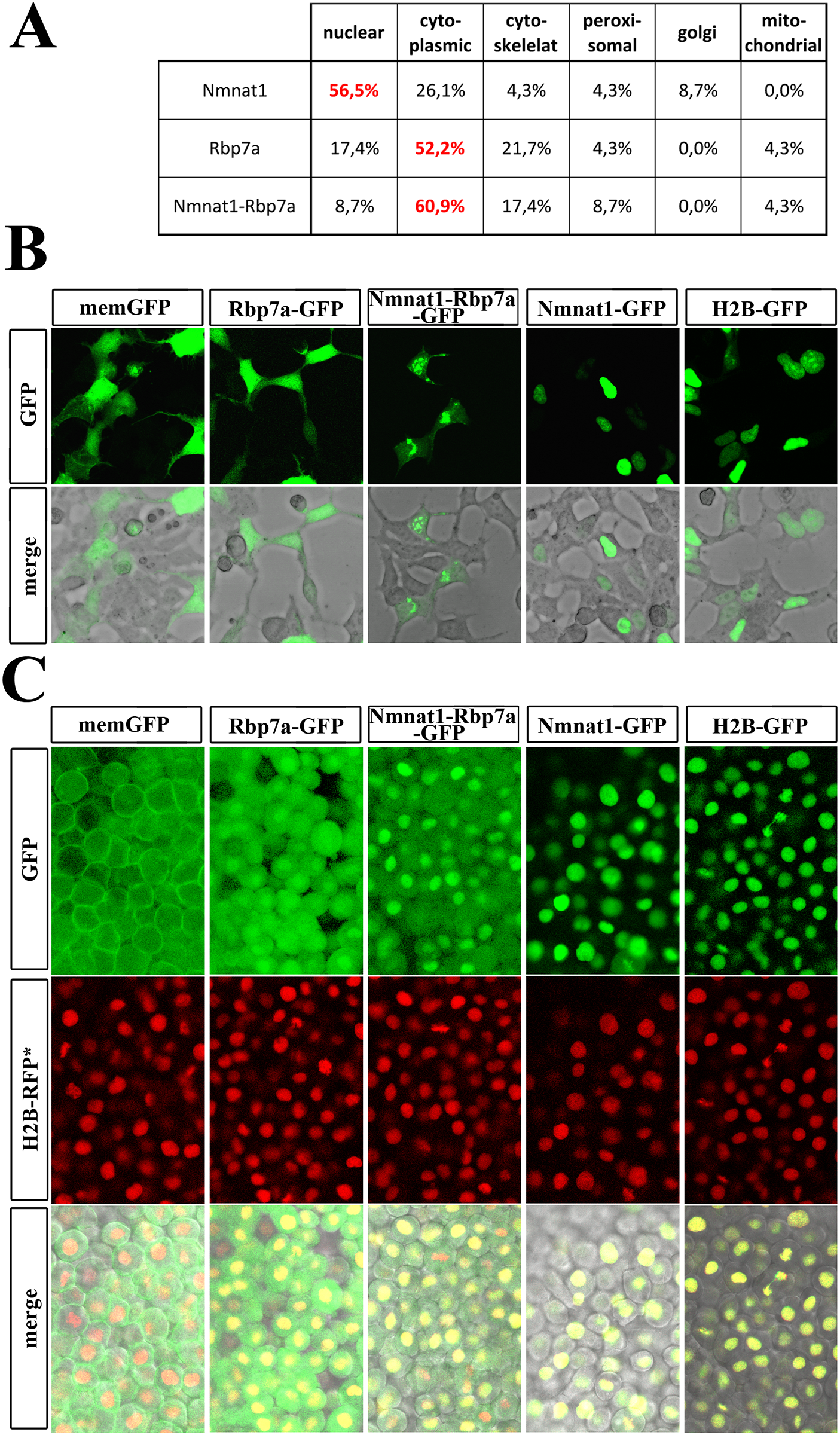
Fig. 5
Subcellular localization of Nmnat1, Rbp7a and Nmnat1-Rbp7a proteins.
[A] PSORT based predictions for sub-cellular location of Nmnat1, Rbp7a and Nmnat1-Rbp7a fusion. [B] Confocal image scans of HEK cells transfected with indicated fusion proteins. Note the strictly cytoplasmic and nuclear GFP-signals for Rbp7a-GFP and Nmnat1-Rbp7a-GFP, respectively. Nmnat1-Rbp7a-GFP transfected cells show weak GFP signals throughout the cytoplasm and stronger focal signals in association with the nuclei and cellular protrusions. [C] Confocal image scans of 6hpf embryo injected with mRNA encoding indicated GFP-tagged proteins. Co-injected mRNA encoding H2B-RFP (nuclear RFP, middle column) was used as loading control and to outline nuclei. Control injections of H2B-GFP and memGFP (membrane GFP) were used to document GFP/RFP co-expression. Rbp7a-GFP and Nmnat1-Rbp7a-GFP both showed nuclear and cytoplasmic localizations; but Nmnat1-GFP was only found in the nucleus.