
|
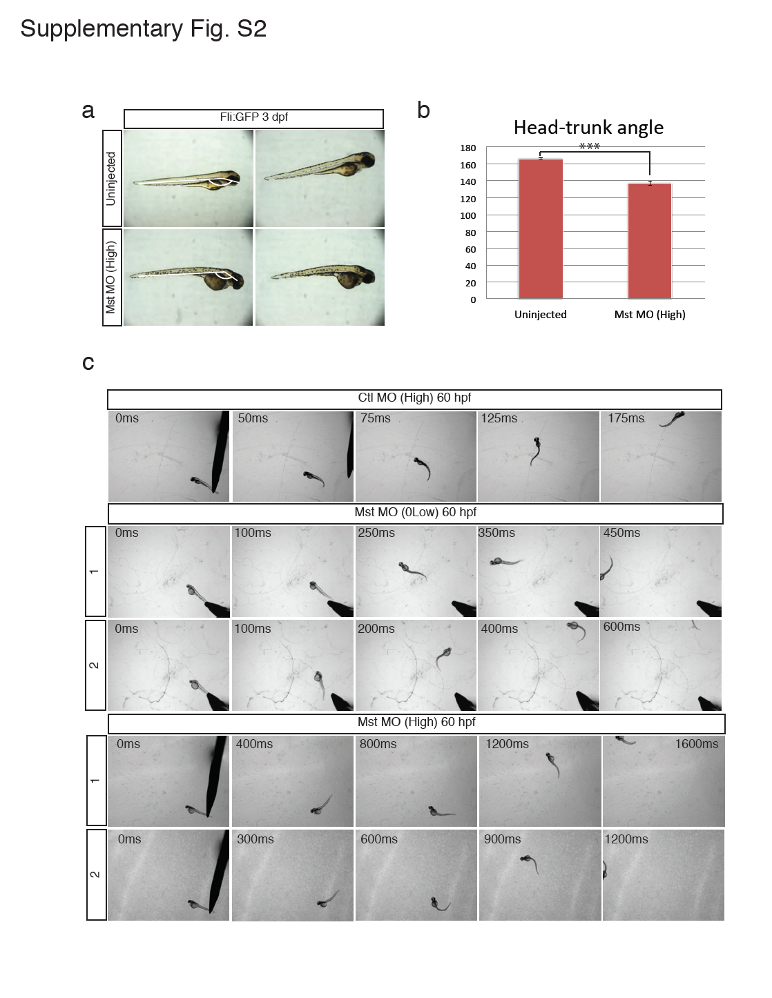
Fig. S2
Morphology and motor function of the mysterin morphant.
(a and b) Observation of mysterin morphants at 3 dpf by stereoscopic microscopy. Development of morphants treated with a high dose of the MO targeting mysterin-α is slightly delayed in comparison with that of control embryos. Control embryos and morphants showed head-trunk angles of 166.4° ± 1.2° (n=14) and 137.1° ± 2.6° (n=10), respectively, suggesting significant delay (approximately 20 hr) in embryogenesis. ***P < 0.001. Animals carry the fli:EGFP transgene, although vascular endothelial cells were not assessed in this figure. (c) Additional successive images of embryonic swimming at 60 hpf. Tracked embryos are independent of those shown in Fig. 1c.