
|
Fig. 6
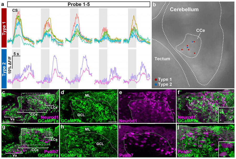
Two types of conditioning-associated neurons. We found two types of conditioning-associated neurons that were activated by the CS. Type I neurons responded quickly to the CS, while type II neurons were activated after a delay. (a) Neuronal activity (ΔF/F) of type I and II neurons of Tg(elavl3:GAL4-VP16); Tg(UAS:GCaMP7a) larvae during the probe session (1st–5th trials), showing data from five type I neurons and two type II neurons. Gray boxes indicate the timing of the CS presentation. (b) Location of the type I (red dots) and II neurons (blue dots) in the CCe. These neurons were located close to each other in the CCe. (c–f) Immunostaining of Tg(elavl3:GAL4-VP16); Tg(UAS:GCaMP7a) about 20-dpf larval brains with anti-GFP (green) and anti-Neurod1 (magenta) antibodies. (d–f) Higher magnification views of the dotted box in (c). The inset in (f) is a higher magnification view of the dotted box in (f). Neurod1 signals mark granule-cell nuclei. Note that Tg(elavl3:GAL4-VP16); Tg(UAS:GCaMP7a) fish expressed GCaMP7a in most of the granule cells in the Va/CCe/LCa (Va: valvula cerebelli) (white asterisks in the inset in f). (g–j) Immunostaining of Tg(elavl3:GAL4-VP16); Tg(UAS:GCaMP7a) brains with anti-GFP (green) and anti-Pvalb7 (magenta) antibodies. (h–j) Higher magnification views of the dotted box in (g). The inset in (j) is a higher magnification view of the dotted box in (j). Pvalb7 signals mark both the neurites (axons and dendrites) and somata of Purkinje cells. No Purkinje cells expressed GCaMP7a (Table S2). Pvalb7+ cells in the GCL are also Purkinje cells as migration of Purkinje cells was not completed at the stage. See Figs 1 and 3 for abbreviations. Scale bars: 50 μm in (c) (applied to g); 20 μm in (d) (applied to d–f, h–j); 10 μm in the inset in (f) (applied to the inset in j).