
|
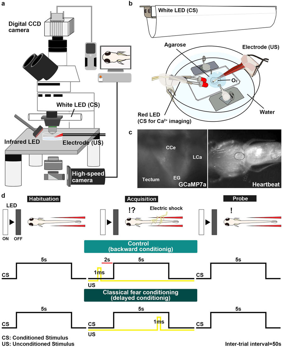
Fig. 1
Method for investigating classical fear conditioning in zebrafish. (a,b) Schematic of the experimental setup for classical fear conditioning using zebrafish larvae at about 20 dpf. The larva was restrained in agarose gel, and O2 gas was provided from below to support respiration. Extinguishment of a white or red LED was given as the CS, and an electric shock was given as the US. (c) Imaging of Ca2+ signals of cerebellar neurons (left panel); and heartbeats (HB, right panel) in Tg(elavl3:GAL4-VP16); Tg(UAS:GCaMP7a) larvae. Images showing the movement of the heart were obtained from underneath with an infrared digital camera. The heart is indicated by a dotted circle. CCe, corpus cerebelli; EG, eminentia granularis; LCa, lobus caudalis cerebelli. (d) Experimental paradigm for delayed classical fear conditioning. In the habituation session, CS alone was given for 5 s per trial; 10–15 trials were performed. In the acquisition session, a paired CS and US (1-ms electric shock given 4 s after the CS onset) was given in each trial; 20 trials were performed. In the probe session, the CS alone was given in each trial and 10 trials were performed. As a control (backward conditioning), the US was given 2 s before the CS onset in the acquisition session. Trials were separated by a 50-s interval. A 50-s interval was also provided between sessions.