
Fig. S3
- ID
- ZDB-IMAGE-171011-20
- Publication
- Nakayama et al., 2017 - Comprehensive analysis of target genes in zebrafish embryos reveals gbx2 involvement in neurogenesis
- All Figures
- Figures for Nakayama et al., 2017
|
Fig. S3
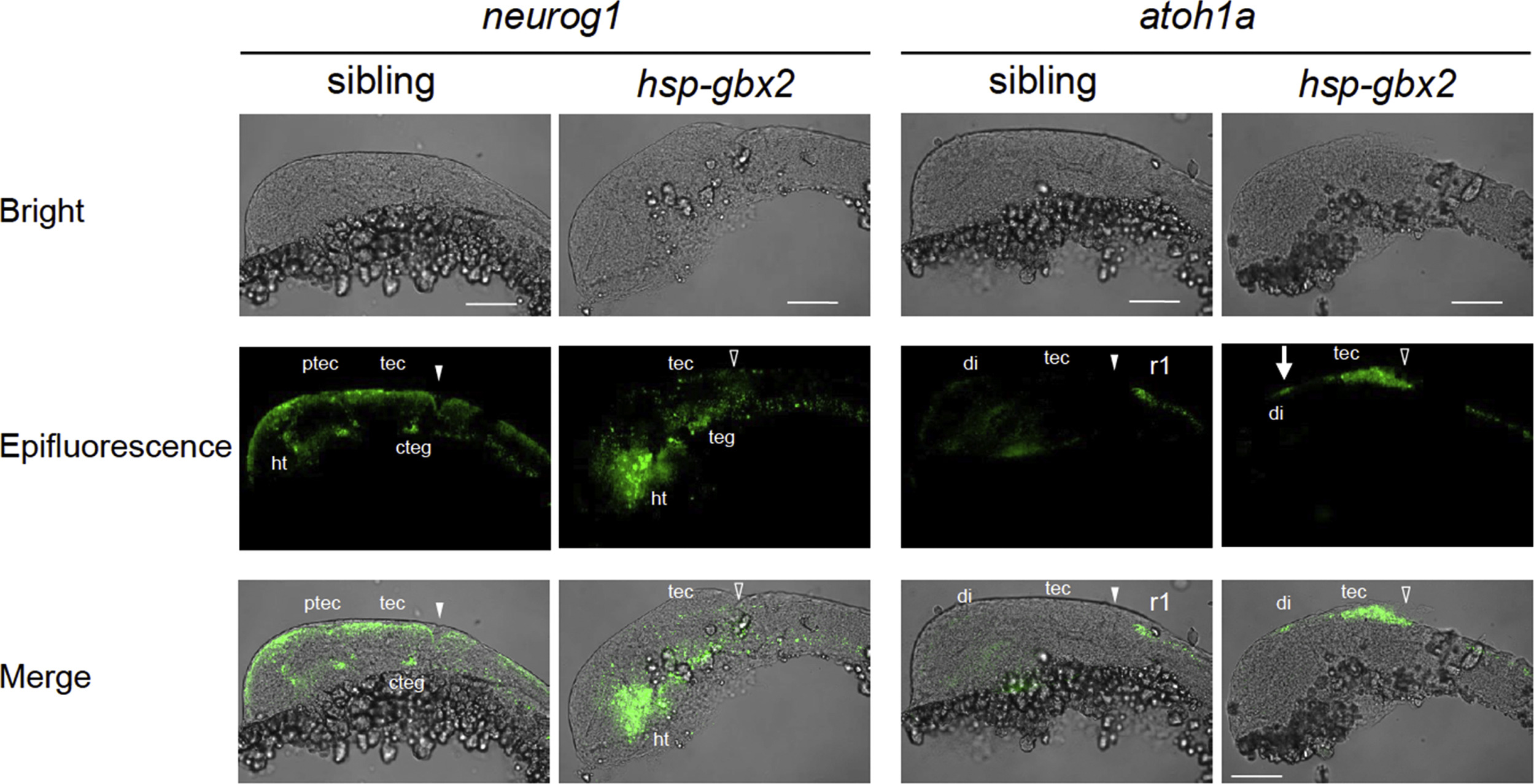
Fluorescent in situ hybridization analysis of the effects of hsp-gbx2 induction on proneural genes. Embryos obtained by mating Tg(hsp70l:gbx2) and wild-type fish were subjected to heat shock at the bud stage and analyzed at 24 hpf for the expression of proneural genes by fluorescent in situ hybridization. The genotypes of the stained embryos were confirmed by PCR. Lateral views with anterior to the left and dorsal to the top are shown. Arrows, ectopic expression in the diencephalon; cteg, caudal tegmentum; di, diencephalon; ht, hypothalamus; ptec, pretectum; r1, rhombomere 1; tec, tectum; teg, tegmentum; solid and open arrowheads, normal and disrupted MHB, respectively. Scale bars, 100 µm.
Reprinted from Developmental Biology, 430(1), Nakayama, Y., Inomata, C., Yuikawa, T., Tsuda, S., Yamasu, K., Comprehensive analysis of target genes in zebrafish embryos reveals gbx2 involvement in neurogenesis, 237-248, Copyright (2017) with permission from Elsevier. Full text @ Dev. Biol.