
Fig. 6
- ID
- ZDB-IMAGE-171011-18
- Genes
- Publication
- Nakayama et al., 2017 - Comprehensive analysis of target genes in zebrafish embryos reveals gbx2 involvement in neurogenesis
- All Figures
- Figures for Nakayama et al., 2017
|
Fig. 6
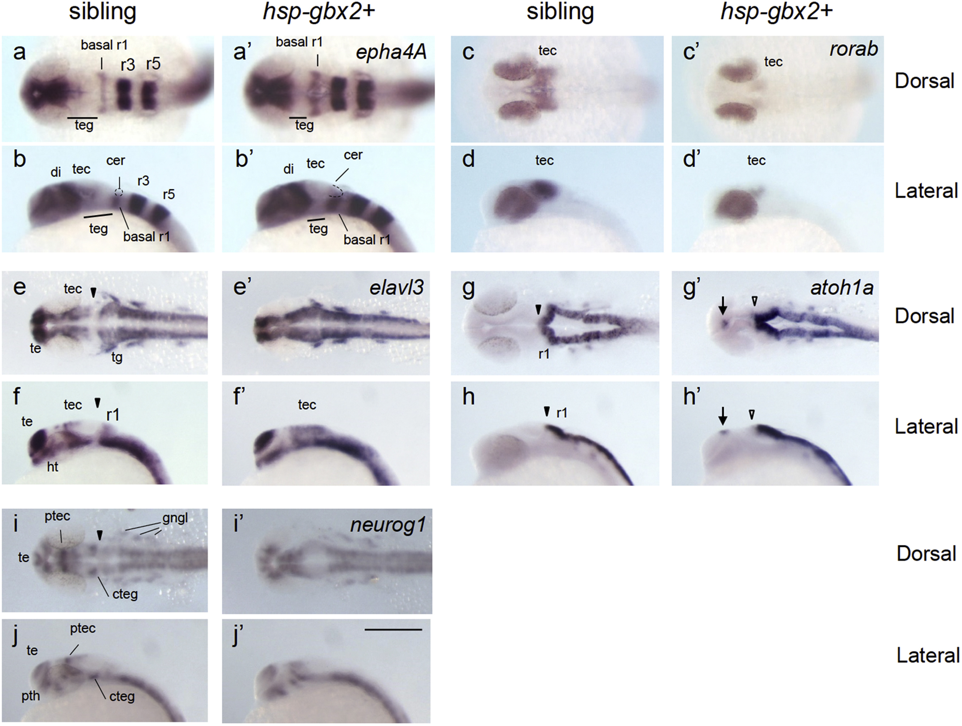
gbx2 promotes neurogenesis in the midbrain-hindbrain region. Embryos obtained by mating Tg(hsp70l:gbx2) and wild-type fish were subjected to heat shock at the bud stage and analyzed at 24 hpf for the expression of neurogenesis genes by WISH. The genotypes of the stained embryos were confirmed by PCR (a–j, sibling; a'–j', hsp-gbx2+). Dorsal views with anterior to the left (a, c, e, g, i, a', c', e', g', i') and lateral views with anterior to the left and dorsal to the top (b, d, f, h, j, b', d', f', h', j') are shown. Arrows, ectopic expression; cer, cerebellum; basal r1, basal region of rhombomere 1; cteg, caudal tegmentum; di, diencephalon; gngl, cranial ganglion; ht, hypothalamus; ptec, pretectum; pth, prethalamus; r1/r3/r5, rhombomere 1/3/5; te, telencephalon; tec, tectum; teg, tegmentum; tg, trigeminal ganglion; black arrowheads, normal MHB; open arrowheads, abnormal MHB. Scale bars, 200 µm.
Reprinted from Developmental Biology, 430(1), Nakayama, Y., Inomata, C., Yuikawa, T., Tsuda, S., Yamasu, K., Comprehensive analysis of target genes in zebrafish embryos reveals gbx2 involvement in neurogenesis, 237-248, Copyright (2017) with permission from Elsevier. Full text @ Dev. Biol.