
|
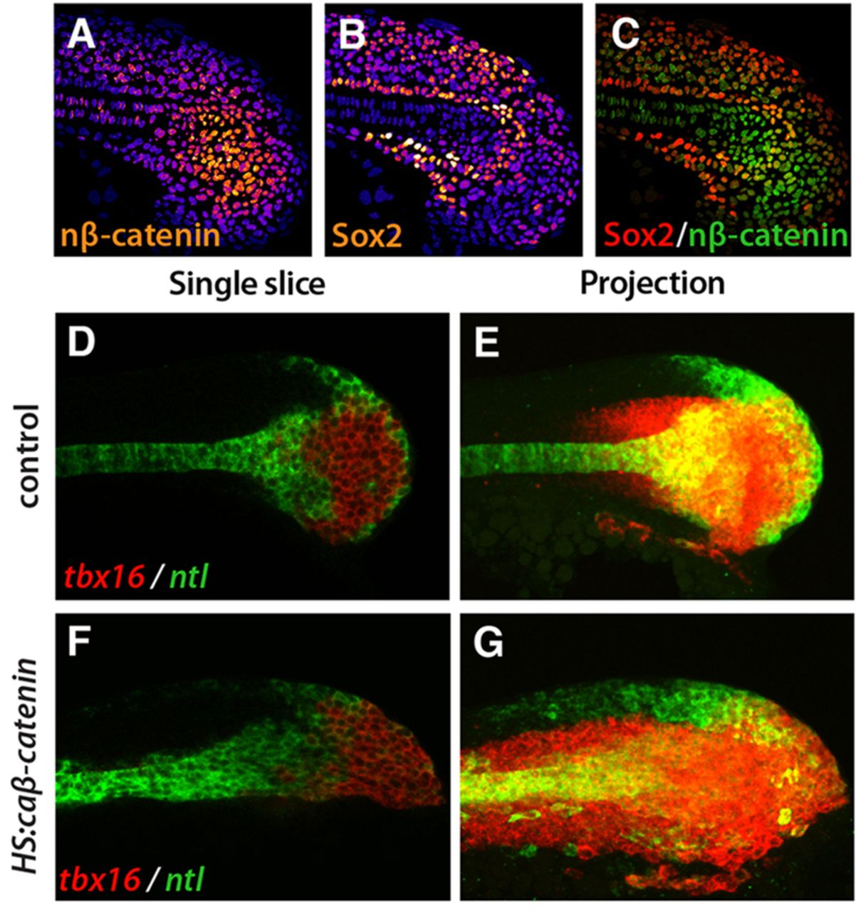
Fig. 4
tbx16 is Wnt responsive. (A-C) Immunofluorescence shows cells in the tailbud with nuclear β-catenin (nβ-catenin; A, orange; cytoplasmic and membrane-bound β-catenin have been eliminated from the image; see Materials and Methods) or Sox2 protein (B, orange). DAPI labels nuclei (blue). The highest levels of nβ-catenin and Sox2 are white and the lowest levels are purple. (C) A composite image shows that there is little overlap between the Sox2 domain (red) and the nuclear β-catenin domain (green). (D-G) Fluorescent whole-mount in situ hybridization shows tbx16 (red) and ntl (green) with HS:caβ-catenin (F,G) and in control (D,E) at 4 h post-heat shock. Single slices (D,F) and maximum intensity projections (E,G) are shown. In all images, anterior is to the left and posterior is to the right.