
Image
|
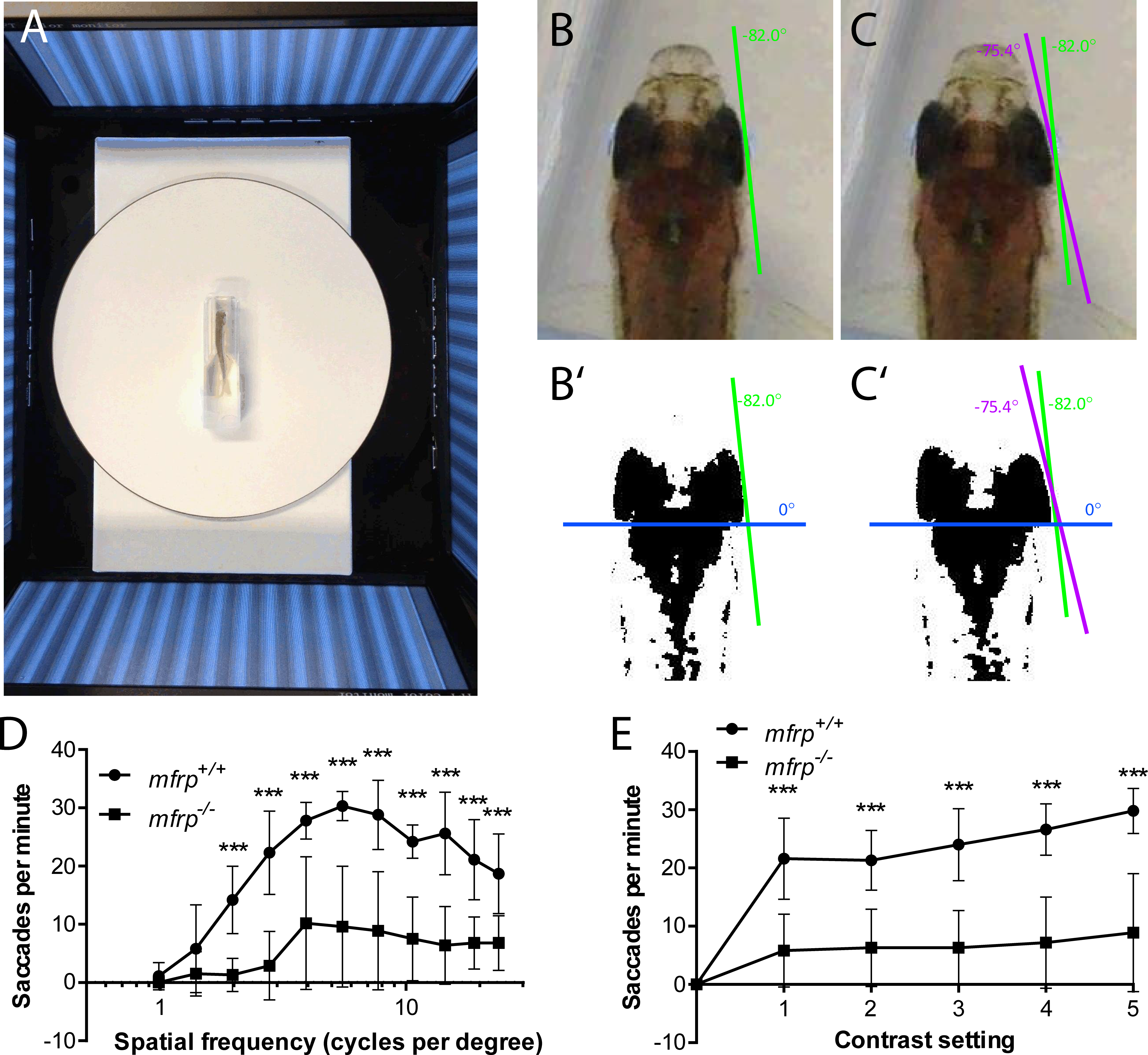
Figure Caption
Fig. 8
Zebrafish mutant for mfrp show reduced optokinetic response. (A) Zebrafish undergoing OKR assay are placed in a plastic cuvette at the center of a testing arena made of four computer monitors that simulate a rotating drum. (B, B', C, C') Images of zebrafish eye positions were taken immediately before (B) and after (C) a rapid eye reset following a smooth pursuit, or saccade. Images were thresholded in ImageJ (http://imagej.nih.gov/ij/; provided in the public domain by the National Institutes of Health, Bethesda, MD, USA) to highlight the highly melanized posterior part of the eye used to define eye position. (D) Fish that were mfrp−/− showed lower visual acuity than mfrp+/+ siblings at 12 mpf by OKR assay. (E) Fish that were mfrp−/− showed fewer saccades per minute than mfrp+/+ siblings by OKR assay when contrast settings were reduced; however, this appeared to be due to reduced visual acuity rather than lack of contrast sensitivity. Graphs show mean saccades per minute ± SD.
Figure Data
Acknowledgments
This image is the copyrighted work of the attributed author or publisher, and
ZFIN has permission only to display this image to its users.
Additional permissions should be obtained from the applicable author or publisher of the image.
Full text @ Invest. Ophthalmol. Vis. Sci.