
Image
|
Figure Caption
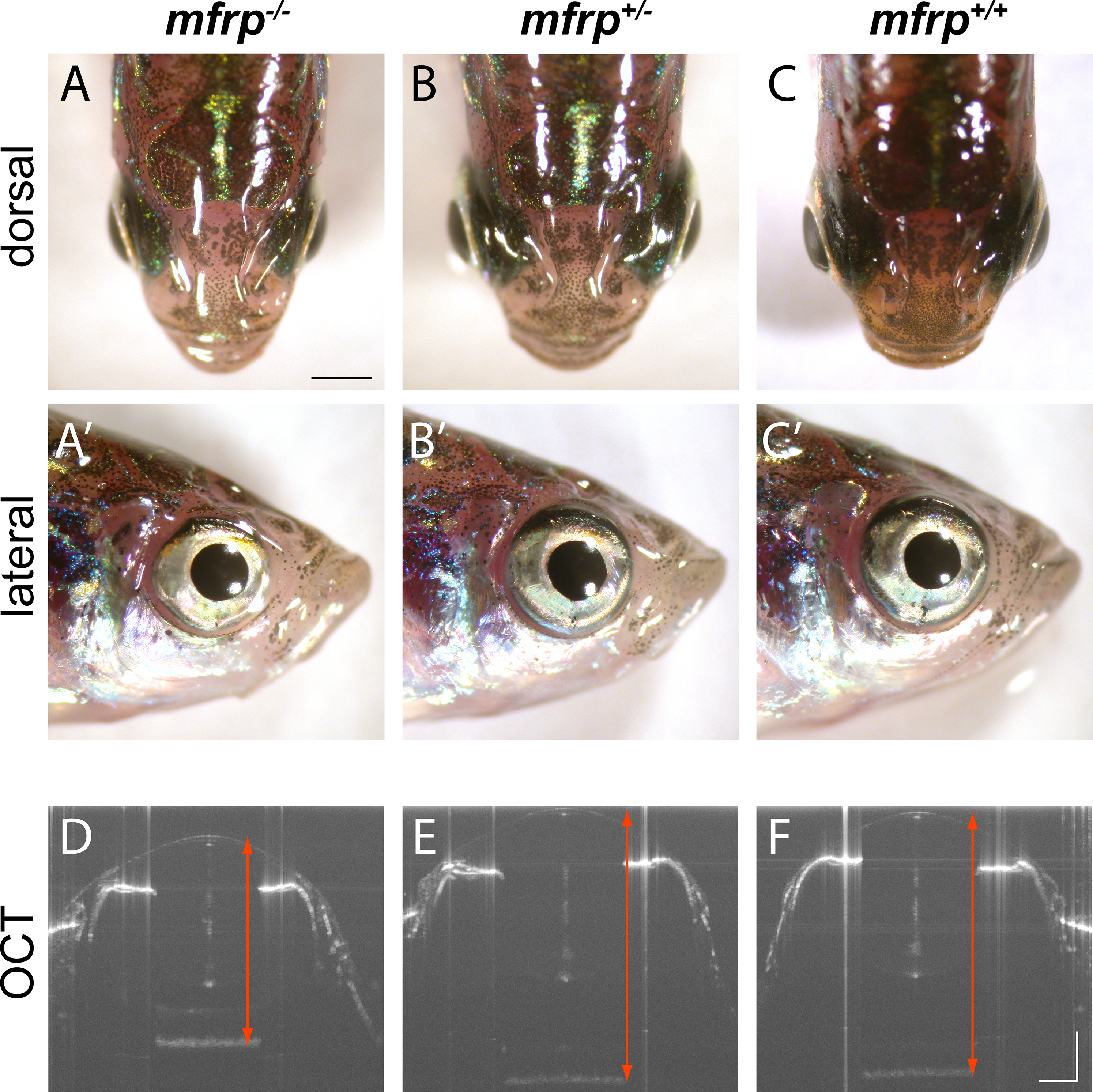
Fig. 3
Zebrafish mfrp mutants show reduced eye size consistent with nanophthalmia. (A, B, C) Dorsal views of mfrp mutant zebrafish and sibling heterozygous and wild-type controls show mfrp homozygotes have smaller eye globes that do not project from the head as much as in their control siblings. (A', B', C') Reduced eye size is also apparent in lateral views. (D, E, F) In vivo SD-OCT imaging shows the reduced axial length (front of cornea to back of RPE; red arrows) associated with homozygous mfrp mutants. Scale bars: (A–C), 1 mm; (D–F), 300 μm.
Figure Data
Acknowledgments
This image is the copyrighted work of the attributed author or publisher, and
ZFIN has permission only to display this image to its users.
Additional permissions should be obtained from the applicable author or publisher of the image.
Full text @ Invest. Ophthalmol. Vis. Sci.