
Image
|
Figure Caption
Fig. 2
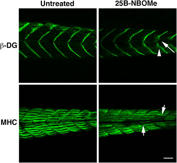
Microscopic immunofluorescent photographs after immunostaining for β-dystroglycan and myosin heavy chain in skeletal muscle of zebrafish larvae with and without treatment by 25B-NBOMe at 0.5 μg/mL for 2 days. The arrowhead and arrow in the right upper panel show irregularity and disruption of the myosepta, respectively. The arrows shown in the right lower panel show myofibril injuries inflicted by 25B-NBOMe. The bar shows the length of 50 μm
Figure Data
Acknowledgments
This image is the copyrighted work of the attributed author or publisher, and
ZFIN has permission only to display this image to its users.
Additional permissions should be obtained from the applicable author or publisher of the image.
Full text @ Forensic Toxicol