
Image
|
Figure Caption
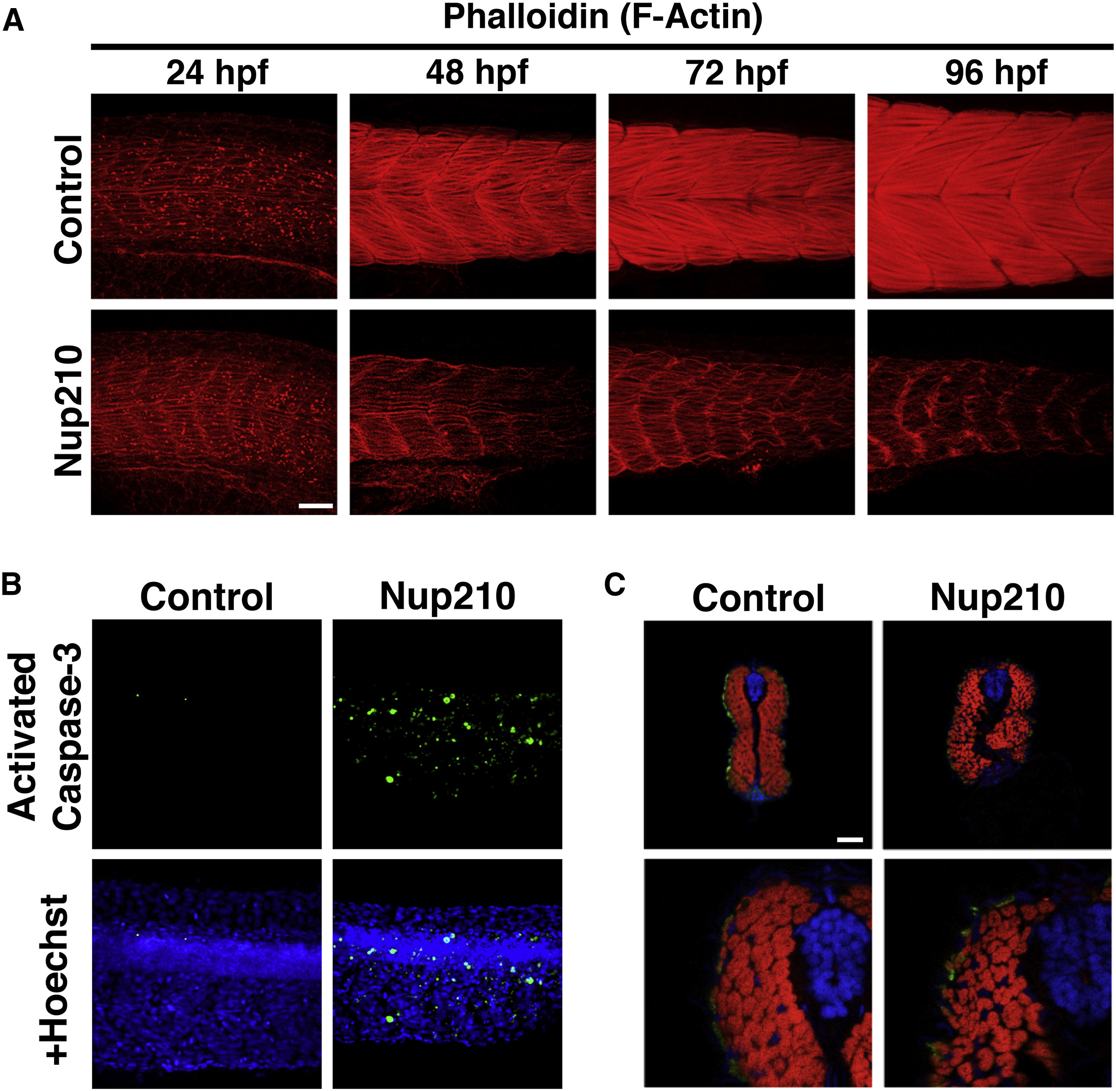
Fig. 3
Nup210 Depletion Inhibits Myofibrillogenesis and Results in Muscle Cell Death
(A) Thin filament structure in control and Nup210 morphants was analyzed by phalloidin staining at different times of development.
(B) Tail muscle was stained for apoptotic death with an anti-activated caspase-3 antibody in control and Nup210-depleted animals.
(C) Cross-sections of zebrafish animals at 96 hpf were stained with the slow muscle marker F59 (green), phalloidin (red), and DAPI (blue) (n = 6–10 embryos).
Representative images of n ≥ 3 independent experiments. n = 10–20 embryos unless otherwise specified. Scale bars, 50 μm.
Figure Data
Acknowledgments
This image is the copyrighted work of the attributed author or publisher, and
ZFIN has permission only to display this image to its users.
Additional permissions should be obtained from the applicable author or publisher of the image.
Reprinted from Developmental Cell, 41, Raices, M., Bukata, L., Sakuma, S., Borlido, J., Hernandez, L.S., Hart, D.O., D'Angelo, M.A., Nuclear Pores Regulate Muscle Development and Maintenance by Assembling a Localized Mef2C Complex, 540-554.e7, Copyright (2017) with permission from Elsevier. Full text @ Dev. Cell