
|
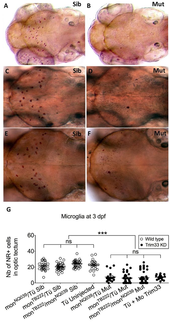
Fig. 1
Moonshine mutants and Trim33 morphants are devoid of microglia. (A–F) In vivo dorsal view of Neutral Red-stained primitive microglia in the midbrain optic tectum of 3-day-old moonshineNQ039/Tü siblings (Sib) (A) and mutants (Mut) (B), moonshineTB222/Tü siblings (C) and mutants (D), and of the siblings (E) and mutants (F) resulting from their complementation cross. (G) Corresponding counts (Nb, number) of Neutral Red-positive (NR+) microglial cells in the midbrain optic tectum of moonshine mutants and siblings, and of Trim33 morphants versus control uninjected embryos. Each dot is one embryo. Error bars show mean±s.e.m. KD, knockdown; Mo, morphant; mon, moonshine; Tü, wild-type Tübingen strain. ***P<0.001; ns, not significant.