
Fig. S5
- ID
- ZDB-IMAGE-170927-21
- Publication
- Madigan et al., 2017 - A Macrophage Response to Mycobacterium leprae Phenolic Glycolipid Initiates Nerve Damage in Leprosy
- All Figures
- Figures for Madigan et al., 2017
|
Fig. S5
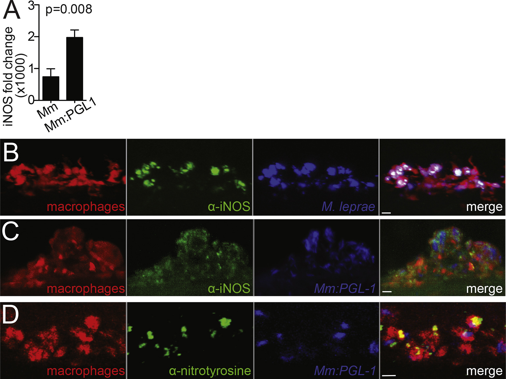
Nitric Oxide Production in Infected Macrophages, Related to Figure 6
(A) Mean (±SEM) fold change of Nos2 (iNOS) transcript in WT murine macrophages, 6 hr after infection with WT M. marinum or M. marinum:PGL-1 (both MOI 1), compared to uninfected cells. (Average of 3 independent experiments; Student’s t test).
(B) Representative confocal images of a macrophage aggregate in an mpeg1 larva infected with M. leprae and stained with α-iNOS antibody. 10 μm bar.
(C) Representative confocal images of a macrophage aggregate in an mpeg1 larva infected with M. marinum:PGL-1 and stained with α-iNOS antibody. 10 μm bar.
(D) Representative confocal images of a macrophage aggregate in larvae as in C, stained with α-nitrotyrosine antibody. 10 μm bar.