
|
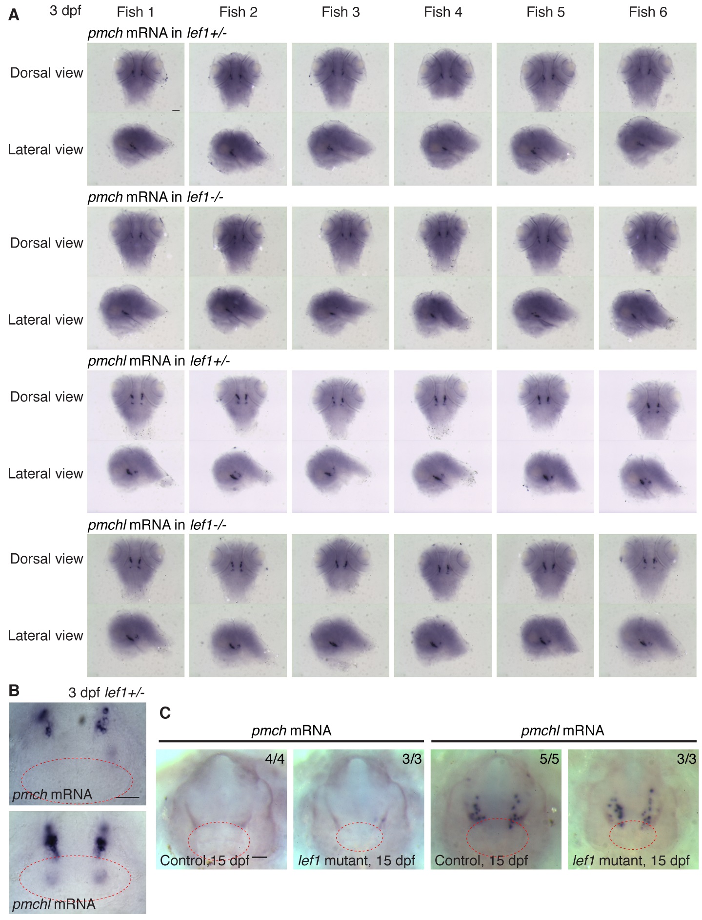
Fig. S6
Normal expression of pmch and pmchl in zebrafish lef1 mutants.
(A-C) Whole mount in situ hybridization images for pmch and pmchl (pmch, like) in the hypothalamus of 3 dpf (A and B) and 15 dpf (C) zebrafish control and lef1 mutant embryos. Images of dorsal views (anterior on top) and lateral views (dorsal on top and anterior on the left) of the same individual lef1+/- or lef1-/- fish were shown in (A). Representative ventral view images of 3 dpf lef1+/- (B), 15 dpf control and lef1 mutant (C) brains centered on the caudal hypothalamus (dashed red outlines) with anterior on top. Number of fish with representative gene expression among total number of fish is labeled on the right upper corner of each image in (C). Scale bar: 100 μm in (A and C); 5 μm in (B).